Fluorine »
PDB 7db8-7e5h »
7dep »
Fluorine in PDB 7dep: S. Aureus Ssbb with 5-Fu
Protein crystallography data
The structure of S. Aureus Ssbb with 5-Fu, PDB code: 7dep
was solved by
E.S.Lin,
Y.H.Huang,
C.Y.Huang,
with X-Ray Crystallography technique. A brief refinement statistics is given in the table below:
| Resolution Low / High (Å) | 28.09 / 3.09 |
| Space group | P 64 2 2 |
| Cell size a, b, c (Å), α, β, γ (°) | 116.962, 116.962, 77.713, 90.00, 90.00, 120.00 |
| R / Rfree (%) | 21.2 / 27.1 |
Fluorine Binding Sites:
The binding sites of Fluorine atom in the S. Aureus Ssbb with 5-Fu
(pdb code 7dep). This binding sites where shown within
5.0 Angstroms radius around Fluorine atom.
In total 2 binding sites of Fluorine where determined in the S. Aureus Ssbb with 5-Fu, PDB code: 7dep:
Jump to Fluorine binding site number: 1; 2;
In total 2 binding sites of Fluorine where determined in the S. Aureus Ssbb with 5-Fu, PDB code: 7dep:
Jump to Fluorine binding site number: 1; 2;
Fluorine binding site 1 out of 2 in 7dep
Go back to
Fluorine binding site 1 out
of 2 in the S. Aureus Ssbb with 5-Fu
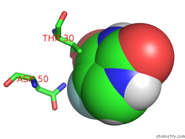
Mono view
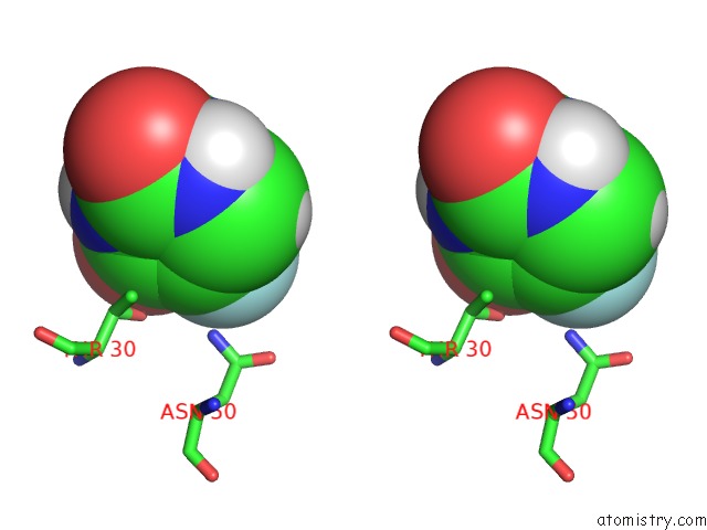
Stereo pair view
Mono view
Stereo pair view
A full contact list of Fluorine with other atoms in the F binding
site number 1 of S. Aureus Ssbb with 5-Fu within 5.0Å range:
|
Fluorine binding site 2 out of 2 in 7dep
Go back to
Fluorine binding site 2 out
of 2 in the S. Aureus Ssbb with 5-Fu
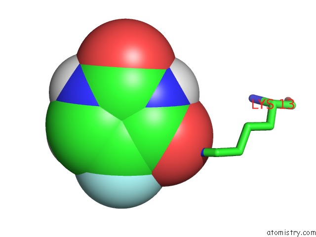
Mono view
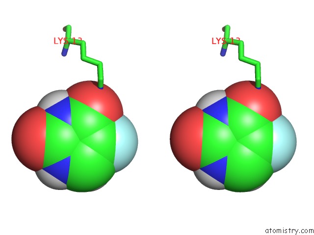
Stereo pair view
Mono view
Stereo pair view
A full contact list of Fluorine with other atoms in the F binding
site number 2 of S. Aureus Ssbb with 5-Fu within 5.0Å range:
|
Reference:
E.S.Lin,
C.Y.Huang.
Crystal Structure of the Single-Stranded Dna-Binding Protein Ssbb in Complex with the Anticancer Drug 5-Fluorouracil: Extension of the 5-Fluorouracil Interactome to Include the Oligonucleotide/Oligosaccharide-Binding Fold Protein Biochem.Biophys.Res.Commun. V. 534 41 2020.
ISSN: ESSN 1090-2104
DOI: 10.1016/J.BBRC.2020.11.125
Page generated: Fri Aug 2 06:27:00 2024
ISSN: ESSN 1090-2104
DOI: 10.1016/J.BBRC.2020.11.125
Last articles
Zn in 9JYWZn in 9IR4
Zn in 9IR3
Zn in 9GMX
Zn in 9GMW
Zn in 9JEJ
Zn in 9ERF
Zn in 9ERE
Zn in 9EGV
Zn in 9EGW