Fluorine »
PDB 7db8-7e5h »
7e5h »
Fluorine in PDB 7e5h: Human Ppar Alpha Ligand Binding Domain in Complex with APHM6 Obtained By Cocrystallization
Protein crystallography data
The structure of Human Ppar Alpha Ligand Binding Domain in Complex with APHM6 Obtained By Cocrystallization, PDB code: 7e5h
was solved by
T.Oyama,
S.Kamata,
I.Ishii,
H.Miyachi,
with X-Ray Crystallography technique. A brief refinement statistics is given in the table below:
| Resolution Low / High (Å) | 42.75 / 1.66 |
| Space group | P 1 21 1 |
| Cell size a, b, c (Å), α, β, γ (°) | 44.599, 61.923, 53.148, 90, 106.54, 90 |
| R / Rfree (%) | 20.3 / 23.9 |
Fluorine Binding Sites:
The binding sites of Fluorine atom in the Human Ppar Alpha Ligand Binding Domain in Complex with APHM6 Obtained By Cocrystallization
(pdb code 7e5h). This binding sites where shown within
5.0 Angstroms radius around Fluorine atom.
In total 2 binding sites of Fluorine where determined in the Human Ppar Alpha Ligand Binding Domain in Complex with APHM6 Obtained By Cocrystallization, PDB code: 7e5h:
Jump to Fluorine binding site number: 1; 2;
In total 2 binding sites of Fluorine where determined in the Human Ppar Alpha Ligand Binding Domain in Complex with APHM6 Obtained By Cocrystallization, PDB code: 7e5h:
Jump to Fluorine binding site number: 1; 2;
Fluorine binding site 1 out of 2 in 7e5h
Go back to
Fluorine binding site 1 out
of 2 in the Human Ppar Alpha Ligand Binding Domain in Complex with APHM6 Obtained By Cocrystallization
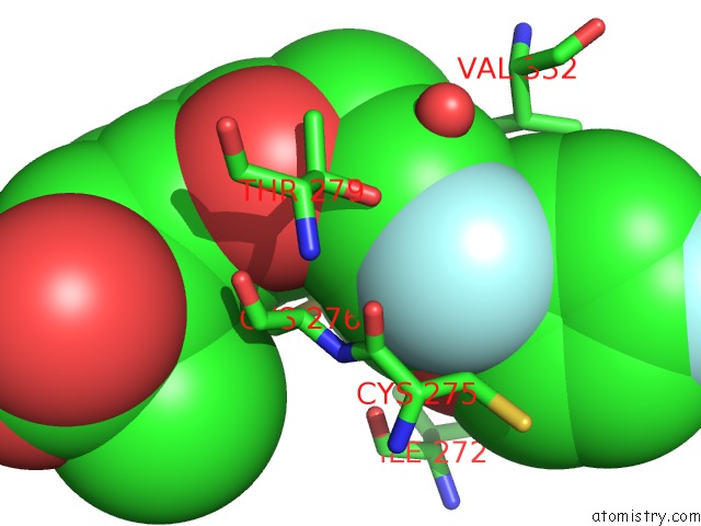
Mono view
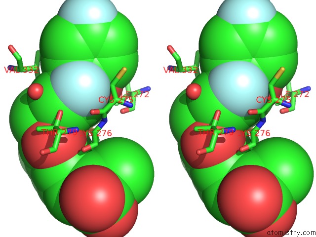
Stereo pair view
Mono view
Stereo pair view
A full contact list of Fluorine with other atoms in the F binding
site number 1 of Human Ppar Alpha Ligand Binding Domain in Complex with APHM6 Obtained By Cocrystallization within 5.0Å range:
|
Fluorine binding site 2 out of 2 in 7e5h
Go back to
Fluorine binding site 2 out
of 2 in the Human Ppar Alpha Ligand Binding Domain in Complex with APHM6 Obtained By Cocrystallization
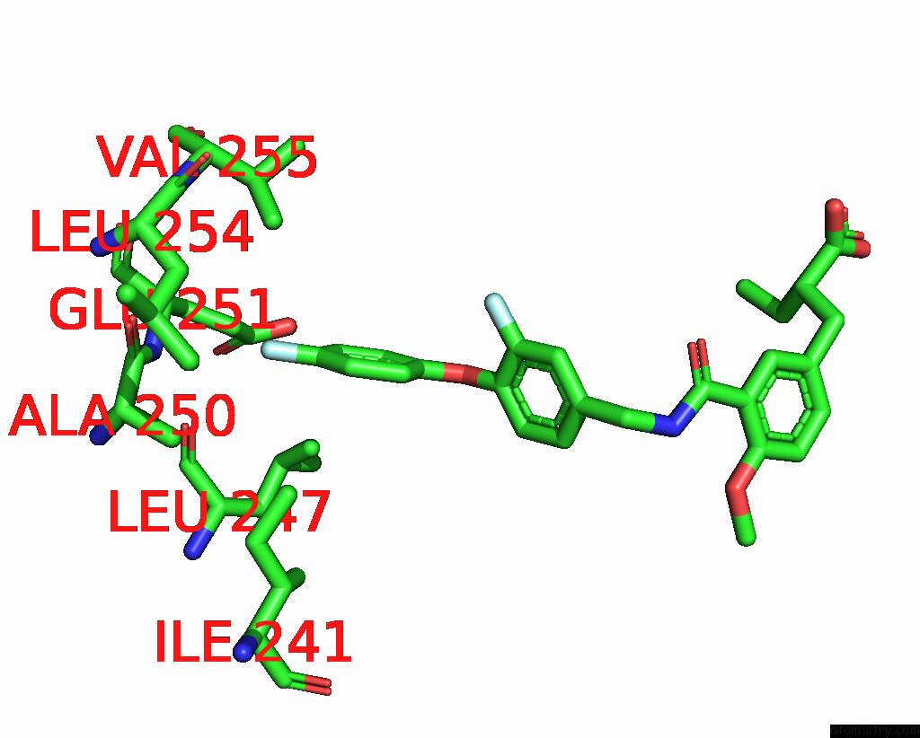
Mono view
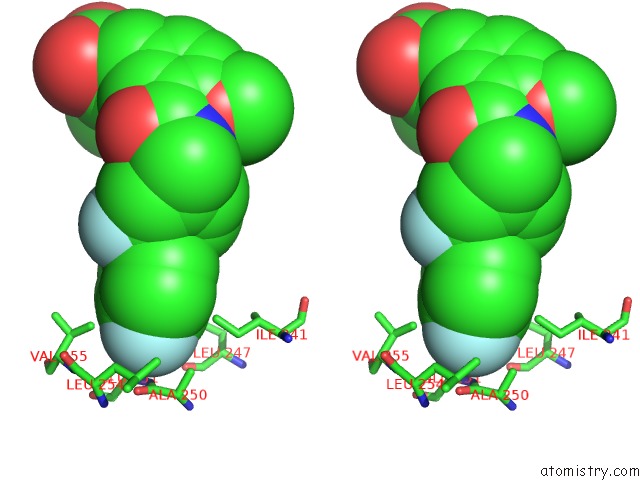
Stereo pair view
Mono view
Stereo pair view
A full contact list of Fluorine with other atoms in the F binding
site number 2 of Human Ppar Alpha Ligand Binding Domain in Complex with APHM6 Obtained By Cocrystallization within 5.0Å range:
|
Reference:
T.Oyama,
S.Kamata,
I.Ishii,
H.Miyachi.
Crystal Structures of the Human Peroxisome Proliferator-Activated Receptor (Ppar) Alpha Ligand-Binding Domain in Complexes with A Series of Phenylpropanoic Acid Derivatives Generated By A Ligand-Exchange Soaking Method. Biol.Pharm.Bull. V. 44 1202 2021.
ISSN: ISSN 0918-6158
PubMed: 34471048
DOI: 10.1248/BPB.B21-00220
Page generated: Fri Aug 2 06:36:52 2024
ISSN: ISSN 0918-6158
PubMed: 34471048
DOI: 10.1248/BPB.B21-00220
Last articles
Zn in 9J0NZn in 9J0O
Zn in 9J0P
Zn in 9FJX
Zn in 9EKB
Zn in 9C0F
Zn in 9CAH
Zn in 9CH0
Zn in 9CH3
Zn in 9CH1