Fluorine in PDB 7erb: Crystal Structure of Human Biliverdin IX-Beta Reductase B with Ataluren (Ptc)
Enzymatic activity of Crystal Structure of Human Biliverdin IX-Beta Reductase B with Ataluren (Ptc)
All present enzymatic activity of Crystal Structure of Human Biliverdin IX-Beta Reductase B with Ataluren (Ptc):
1.3.1.24;
1.5.1.30;
Protein crystallography data
The structure of Crystal Structure of Human Biliverdin IX-Beta Reductase B with Ataluren (Ptc), PDB code: 7erb
was solved by
C.Griesinger,
D.Lee,
K.S.Ryu,
M.Kim,
J.H.Ha,
with X-Ray Crystallography technique. A brief refinement statistics is given in the table below:
Resolution Low / High (Å)
|
41.71 /
1.50
|
Space group
|
P 21 21 21
|
Cell size a, b, c (Å), α, β, γ (°)
|
83.423,
117.338,
82.045,
90,
90,
90
|
R / Rfree (%)
|
17.1 /
19.9
|
Fluorine Binding Sites:
The binding sites of Fluorine atom in the Crystal Structure of Human Biliverdin IX-Beta Reductase B with Ataluren (Ptc)
(pdb code 7erb). This binding sites where shown within
5.0 Angstroms radius around Fluorine atom.
In total 4 binding sites of Fluorine where determined in the
Crystal Structure of Human Biliverdin IX-Beta Reductase B with Ataluren (Ptc), PDB code: 7erb:
Jump to Fluorine binding site number:
1;
2;
3;
4;
Fluorine binding site 1 out
of 4 in 7erb
Go back to
Fluorine Binding Sites List in 7erb
Fluorine binding site 1 out
of 4 in the Crystal Structure of Human Biliverdin IX-Beta Reductase B with Ataluren (Ptc)
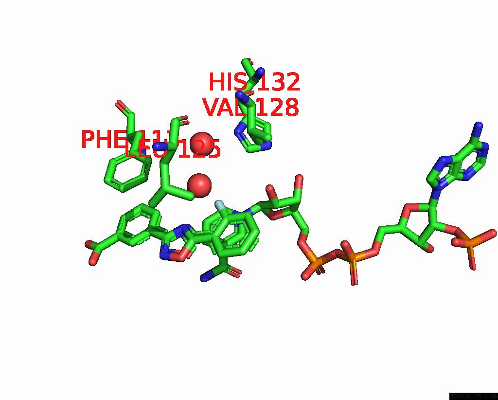
Mono view
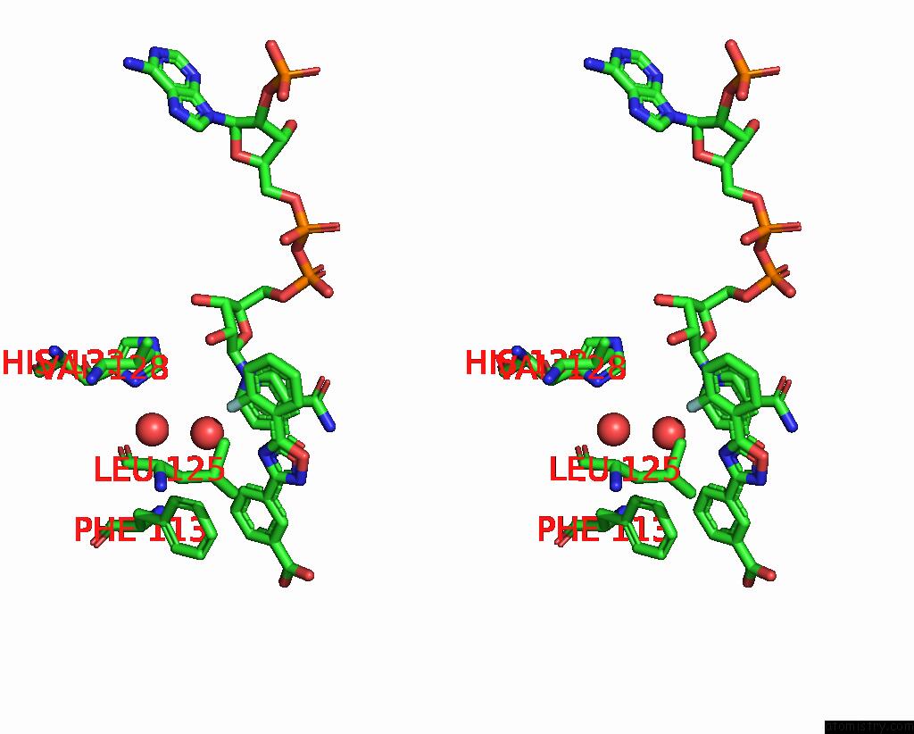
Stereo pair view
|
A full contact list of Fluorine with other atoms in the F binding
site number 1 of Crystal Structure of Human Biliverdin IX-Beta Reductase B with Ataluren (Ptc) within 5.0Å range:
probe
|
atom
|
residue
|
distance (Å)
|
B
|
Occ
|
A:F402
b:29.1
occ:1.00
|
F01
|
A:JBF402
|
0.0
|
29.1
|
1.0
|
C02
|
A:JBF402
|
1.4
|
27.3
|
1.0
|
C07
|
A:JBF402
|
2.3
|
25.1
|
1.0
|
C03
|
A:JBF402
|
2.4
|
27.1
|
1.0
|
O
|
A:HOH607
|
2.5
|
18.4
|
1.0
|
N09
|
A:JBF402
|
2.6
|
20.3
|
1.0
|
C08
|
A:JBF402
|
2.7
|
21.5
|
1.0
|
C06
|
A:JBF402
|
3.6
|
25.7
|
1.0
|
C2N
|
A:NAP401
|
3.6
|
7.6
|
1.0
|
C04
|
A:JBF402
|
3.6
|
26.4
|
1.0
|
O2D
|
A:NAP401
|
3.7
|
9.0
|
1.0
|
C3N
|
A:NAP401
|
3.8
|
9.0
|
1.0
|
N1N
|
A:NAP401
|
3.8
|
7.3
|
1.0
|
C10
|
A:JBF402
|
4.0
|
19.9
|
1.0
|
O21
|
A:JBF402
|
4.1
|
21.1
|
1.0
|
C05
|
A:JBF402
|
4.1
|
26.0
|
1.0
|
O
|
A:HOH577
|
4.1
|
17.7
|
1.0
|
C4N
|
A:NAP401
|
4.2
|
8.9
|
1.0
|
C6N
|
A:NAP401
|
4.2
|
8.1
|
1.0
|
CD2
|
A:LEU125
|
4.3
|
17.8
|
1.0
|
C5N
|
A:NAP401
|
4.3
|
8.4
|
1.0
|
C7N
|
A:NAP401
|
4.4
|
10.0
|
1.0
|
C2D
|
A:NAP401
|
4.4
|
7.6
|
1.0
|
C1D
|
A:NAP401
|
4.5
|
7.5
|
1.0
|
N20
|
A:JBF402
|
4.6
|
20.8
|
1.0
|
CG1
|
A:VAL128
|
4.6
|
11.9
|
1.0
|
O7N
|
A:NAP401
|
4.7
|
11.9
|
1.0
|
CE1
|
A:HIS132
|
4.7
|
7.3
|
1.0
|
CD1
|
A:LEU125
|
4.8
|
17.0
|
1.0
|
C19
|
A:JBF402
|
4.8
|
16.1
|
1.0
|
CE2
|
A:PHE113
|
4.9
|
11.6
|
1.0
|
C11
|
A:JBF402
|
4.9
|
17.4
|
1.0
|
|
Fluorine binding site 2 out
of 4 in 7erb
Go back to
Fluorine Binding Sites List in 7erb
Fluorine binding site 2 out
of 4 in the Crystal Structure of Human Biliverdin IX-Beta Reductase B with Ataluren (Ptc)
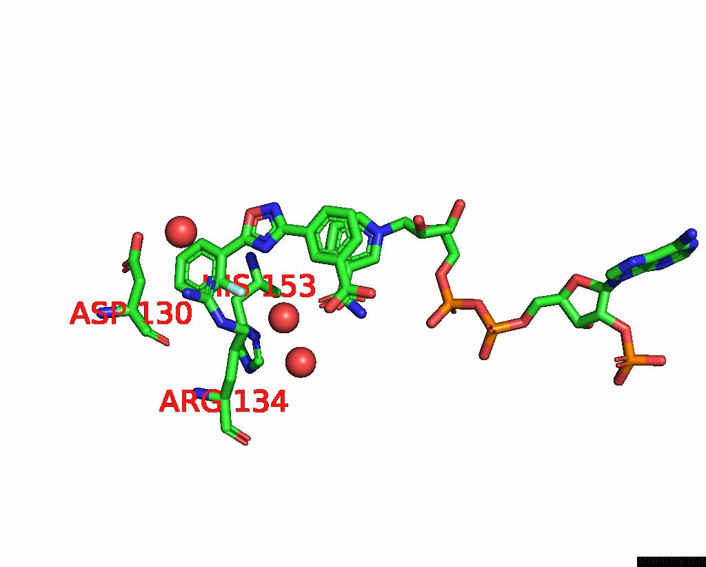
Mono view
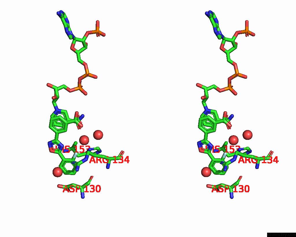
Stereo pair view
|
A full contact list of Fluorine with other atoms in the F binding
site number 2 of Crystal Structure of Human Biliverdin IX-Beta Reductase B with Ataluren (Ptc) within 5.0Å range:
probe
|
atom
|
residue
|
distance (Å)
|
B
|
Occ
|
A:F403
b:57.4
occ:1.00
|
F01
|
A:JBF403
|
0.0
|
57.4
|
1.0
|
C02
|
A:JBF403
|
1.4
|
57.2
|
1.0
|
C07
|
A:JBF403
|
2.4
|
55.2
|
1.0
|
C03
|
A:JBF403
|
2.4
|
58.1
|
1.0
|
O
|
B:HOH525
|
2.5
|
31.2
|
1.0
|
N09
|
A:JBF403
|
2.6
|
51.2
|
1.0
|
C08
|
A:JBF403
|
2.8
|
51.8
|
1.0
|
NH1
|
A:ARG134
|
3.0
|
12.5
|
1.0
|
CD
|
A:ARG134
|
3.3
|
11.3
|
1.0
|
CG
|
A:ARG134
|
3.3
|
9.4
|
1.0
|
ND1
|
B:HIS153
|
3.4
|
18.1
|
1.0
|
C04
|
A:JBF403
|
3.7
|
57.9
|
1.0
|
C06
|
A:JBF403
|
3.7
|
56.2
|
1.0
|
CB
|
B:HIS153
|
3.8
|
17.8
|
1.0
|
C10
|
A:JBF403
|
3.8
|
50.4
|
1.0
|
CZ
|
A:ARG134
|
3.9
|
11.7
|
1.0
|
NE
|
A:ARG134
|
4.0
|
10.8
|
1.0
|
CA
|
B:HIS153
|
4.0
|
16.8
|
1.0
|
CG
|
B:HIS153
|
4.1
|
18.3
|
1.0
|
C05
|
A:JBF403
|
4.1
|
57.2
|
1.0
|
O21
|
A:JBF403
|
4.1
|
51.2
|
1.0
|
C12
|
A:JBF403
|
4.4
|
45.1
|
1.0
|
CE1
|
B:HIS153
|
4.5
|
18.7
|
1.0
|
O7N
|
B:NAP401
|
4.5
|
19.7
|
1.0
|
N
|
B:HIS153
|
4.5
|
18.0
|
1.0
|
O
|
A:ASP130
|
4.6
|
8.5
|
1.0
|
N20
|
A:JBF403
|
4.6
|
51.0
|
1.0
|
C11
|
A:JBF403
|
4.7
|
46.6
|
1.0
|
O
|
B:HOH668
|
4.7
|
47.5
|
1.0
|
O
|
A:HOH611
|
4.8
|
18.3
|
1.0
|
CB
|
A:ARG134
|
4.8
|
8.1
|
1.0
|
|
Fluorine binding site 3 out
of 4 in 7erb
Go back to
Fluorine Binding Sites List in 7erb
Fluorine binding site 3 out
of 4 in the Crystal Structure of Human Biliverdin IX-Beta Reductase B with Ataluren (Ptc)
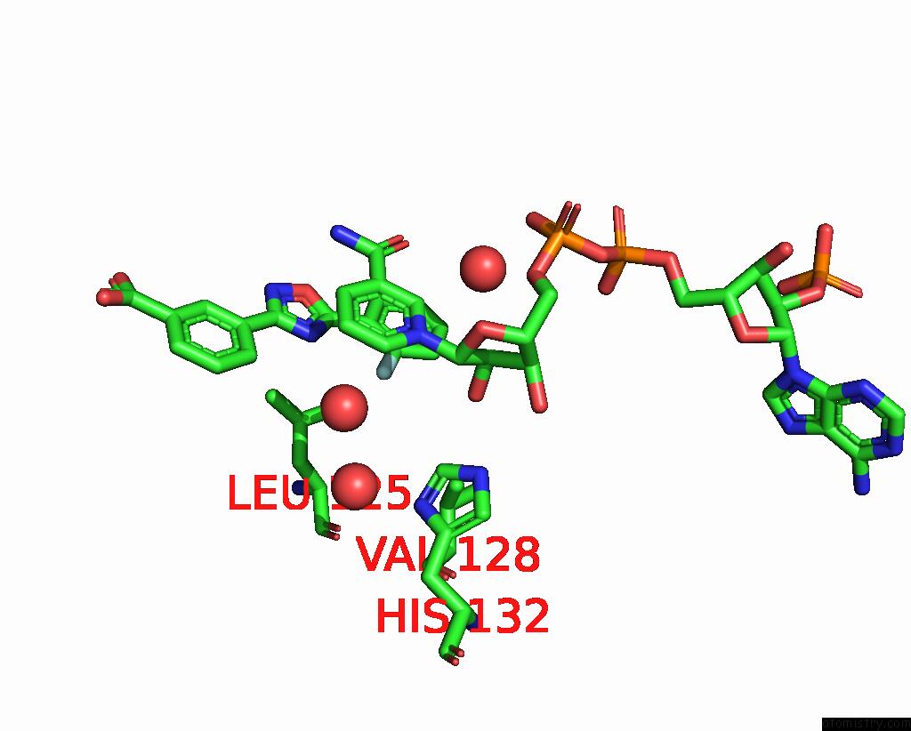
Mono view
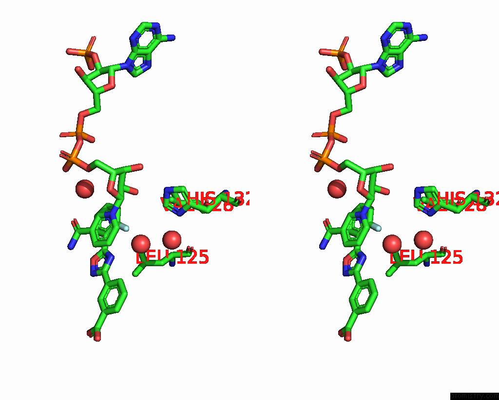
Stereo pair view
|
A full contact list of Fluorine with other atoms in the F binding
site number 3 of Crystal Structure of Human Biliverdin IX-Beta Reductase B with Ataluren (Ptc) within 5.0Å range:
probe
|
atom
|
residue
|
distance (Å)
|
B
|
Occ
|
C:F402
b:24.1
occ:1.00
|
F01
|
C:JBF402
|
0.0
|
24.1
|
1.0
|
C02
|
C:JBF402
|
1.3
|
21.6
|
1.0
|
C07
|
C:JBF402
|
2.3
|
19.3
|
1.0
|
C03
|
C:JBF402
|
2.4
|
20.6
|
1.0
|
O
|
C:HOH619
|
2.5
|
16.6
|
1.0
|
N09
|
C:JBF402
|
2.7
|
17.5
|
1.0
|
C08
|
C:JBF402
|
2.7
|
18.3
|
1.0
|
C2N
|
C:NAP401
|
3.4
|
8.7
|
1.0
|
O2D
|
C:NAP401
|
3.5
|
7.7
|
1.0
|
C06
|
C:JBF402
|
3.5
|
19.5
|
1.0
|
C04
|
C:JBF402
|
3.6
|
19.8
|
1.0
|
C3N
|
C:NAP401
|
3.6
|
8.7
|
1.0
|
N1N
|
C:NAP401
|
3.7
|
7.5
|
1.0
|
C05
|
C:JBF402
|
4.0
|
19.4
|
1.0
|
O21
|
C:JBF402
|
4.1
|
18.7
|
1.0
|
C10
|
C:JBF402
|
4.1
|
17.5
|
1.0
|
C6N
|
C:NAP401
|
4.1
|
8.5
|
1.0
|
C4N
|
C:NAP401
|
4.1
|
9.1
|
1.0
|
C7N
|
C:NAP401
|
4.2
|
10.2
|
1.0
|
O
|
C:HOH578
|
4.2
|
16.9
|
1.0
|
C2D
|
C:NAP401
|
4.2
|
7.1
|
1.0
|
C1D
|
C:NAP401
|
4.3
|
7.6
|
1.0
|
CD2
|
C:LEU125
|
4.3
|
16.4
|
1.0
|
C5N
|
C:NAP401
|
4.4
|
8.0
|
1.0
|
O7N
|
C:NAP401
|
4.4
|
13.4
|
1.0
|
CG1
|
C:VAL128
|
4.6
|
12.0
|
1.0
|
CE1
|
C:HIS132
|
4.7
|
7.9
|
1.0
|
N20
|
C:JBF402
|
4.7
|
17.6
|
1.0
|
O
|
C:HOH540
|
4.8
|
9.5
|
1.0
|
CD1
|
C:LEU125
|
4.9
|
16.3
|
1.0
|
N7N
|
C:NAP401
|
5.0
|
8.7
|
1.0
|
|
Fluorine binding site 4 out
of 4 in 7erb
Go back to
Fluorine Binding Sites List in 7erb
Fluorine binding site 4 out
of 4 in the Crystal Structure of Human Biliverdin IX-Beta Reductase B with Ataluren (Ptc)
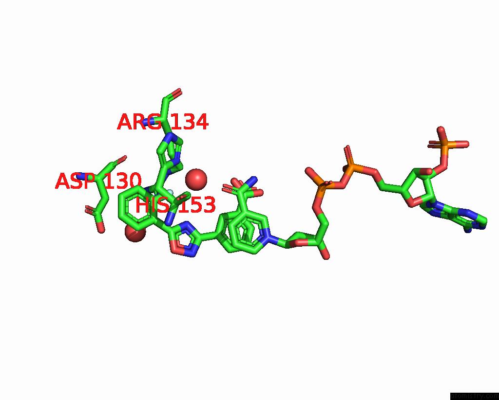
Mono view
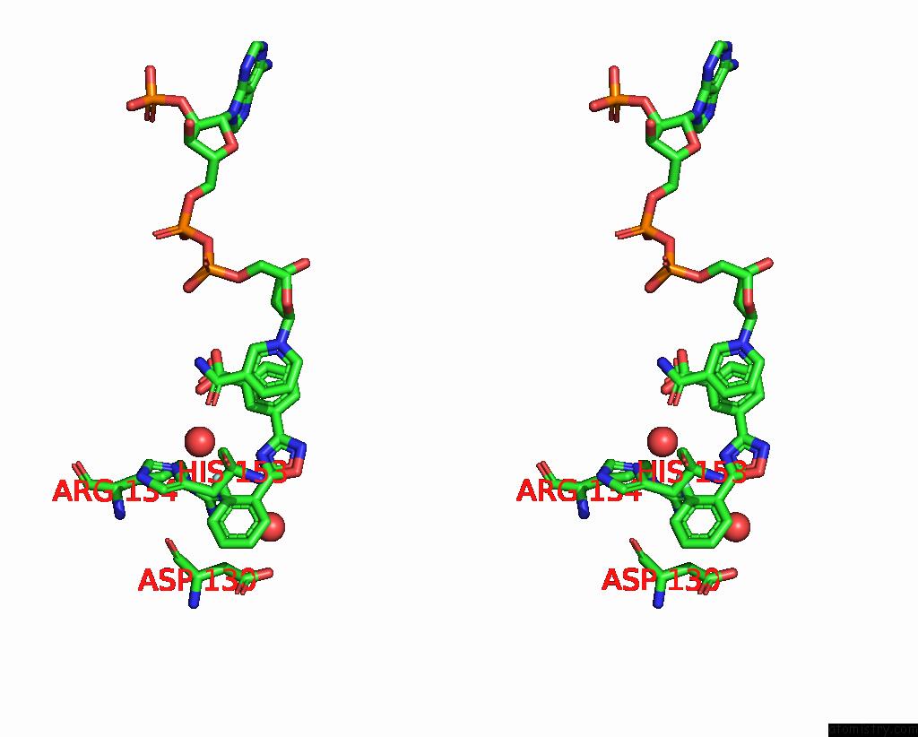
Stereo pair view
|
A full contact list of Fluorine with other atoms in the F binding
site number 4 of Crystal Structure of Human Biliverdin IX-Beta Reductase B with Ataluren (Ptc) within 5.0Å range:
probe
|
atom
|
residue
|
distance (Å)
|
B
|
Occ
|
C:F403
b:54.3
occ:1.00
|
F01
|
C:JBF403
|
0.0
|
54.3
|
1.0
|
C02
|
C:JBF403
|
1.4
|
54.2
|
1.0
|
O
|
D:HOH515
|
2.3
|
29.2
|
1.0
|
C07
|
C:JBF403
|
2.4
|
52.6
|
1.0
|
C03
|
C:JBF403
|
2.4
|
54.6
|
1.0
|
N09
|
C:JBF403
|
2.6
|
48.5
|
1.0
|
C08
|
C:JBF403
|
2.8
|
49.1
|
1.0
|
NH1
|
C:ARG134
|
2.9
|
11.9
|
1.0
|
CG
|
C:ARG134
|
3.3
|
7.6
|
1.0
|
CD
|
C:ARG134
|
3.3
|
9.3
|
1.0
|
ND1
|
D:HIS153
|
3.6
|
18.1
|
1.0
|
C04
|
C:JBF403
|
3.7
|
54.2
|
1.0
|
C06
|
C:JBF403
|
3.7
|
53.5
|
1.0
|
CZ
|
C:ARG134
|
3.8
|
10.7
|
1.0
|
C10
|
C:JBF403
|
3.9
|
47.8
|
1.0
|
NE
|
C:ARG134
|
4.0
|
10.2
|
1.0
|
CB
|
D:HIS153
|
4.0
|
18.6
|
1.0
|
C05
|
C:JBF403
|
4.1
|
53.9
|
1.0
|
O21
|
C:JBF403
|
4.2
|
48.5
|
1.0
|
CG
|
D:HIS153
|
4.2
|
18.5
|
1.0
|
CA
|
D:HIS153
|
4.3
|
17.4
|
1.0
|
C12
|
C:JBF403
|
4.4
|
43.5
|
1.0
|
O
|
C:ASP130
|
4.4
|
9.1
|
1.0
|
O7N
|
D:NAP401
|
4.5
|
20.1
|
1.0
|
O
|
C:HOH618
|
4.6
|
17.7
|
1.0
|
N20
|
C:JBF403
|
4.6
|
48.4
|
1.0
|
C11
|
C:JBF403
|
4.7
|
44.6
|
1.0
|
CE1
|
D:HIS153
|
4.7
|
18.6
|
1.0
|
CB
|
C:ARG134
|
4.8
|
7.0
|
1.0
|
N
|
D:HIS153
|
4.8
|
18.0
|
1.0
|
CB
|
C:ASP130
|
5.0
|
9.9
|
1.0
|
|
Reference:
M.Kim,
J.H.Ha,
J.Choi,
B.R.Kim,
V.Gapsys,
K.O.Lee,
J.G.Jee,
K.S.Chakrabarti,
B.L.De Groot,
C.Griesinger,
K.S.Ryu,
D.Lee.
Repositioning Food and Drug Administration-Approved Drugs For Inhibiting Biliverdin IX Beta Reductase B As A Novel Thrombocytopenia Therapeutic Target. J.Med.Chem. V. 65 2548 2022.
ISSN: ISSN 0022-2623
PubMed: 34957824
DOI: 10.1021/ACS.JMEDCHEM.1C01664
Page generated: Fri Aug 2 06:45:24 2024
|