Fluorine in PDB 7evy: Cryo-Em Structure of Siponimod -Bound Sphingosine-1-Phosphate Receptor 1 in Complex with Gi Protein
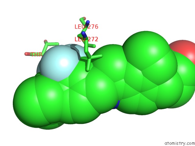
Fluorine Binding Sites:
The binding sites of Fluorine atom in the Cryo-Em Structure of Siponimod -Bound Sphingosine-1-Phosphate Receptor 1 in Complex with Gi Protein
(pdb code 7evy). This binding sites where shown within
5.0 Angstroms radius around Fluorine atom.
In total 3 binding sites of Fluorine where determined in the
Cryo-Em Structure of Siponimod -Bound Sphingosine-1-Phosphate Receptor 1 in Complex with Gi Protein, PDB code: 7evy:
Jump to Fluorine binding site number:
1;
2;
3;
Fluorine binding site 1 out
of 3 in 7evy
Go back to
Fluorine Binding Sites List in 7evy
Fluorine binding site 1 out
of 3 in the Cryo-Em Structure of Siponimod -Bound Sphingosine-1-Phosphate Receptor 1 in Complex with Gi Protein
Mono view
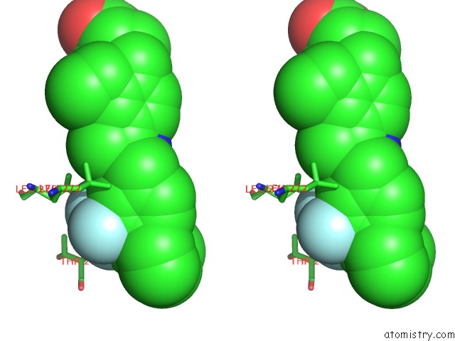
Stereo pair view
|
A full contact list of Fluorine with other atoms in the F binding
site number 1 of Cryo-Em Structure of Siponimod -Bound Sphingosine-1-Phosphate Receptor 1 in Complex with Gi Protein within 5.0Å range:
probe
|
atom
|
residue
|
distance (Å)
|
B
|
Occ
|
D:F401
b:124.3
occ:1.00
|
F26
|
D:J8C401
|
0.0
|
124.3
|
1.0
|
C25
|
D:J8C401
|
1.4
|
124.3
|
1.0
|
CD2
|
D:LEU276
|
2.2
|
126.4
|
1.0
|
F28
|
D:J8C401
|
2.2
|
124.3
|
1.0
|
F27
|
D:J8C401
|
2.2
|
124.3
|
1.0
|
C15
|
D:J8C401
|
2.3
|
124.3
|
1.0
|
C14
|
D:J8C401
|
2.7
|
124.3
|
1.0
|
CG
|
D:LEU276
|
3.5
|
126.4
|
1.0
|
C16
|
D:J8C401
|
3.6
|
124.3
|
1.0
|
CD1
|
D:LEU276
|
4.0
|
126.4
|
1.0
|
C13
|
D:J8C401
|
4.0
|
124.3
|
1.0
|
CG2
|
D:THR207
|
4.1
|
122.5
|
1.0
|
CB
|
D:LEU276
|
4.2
|
126.4
|
1.0
|
C19
|
D:J8C401
|
4.3
|
124.3
|
1.0
|
CB
|
D:CYS206
|
4.5
|
124.1
|
1.0
|
N
|
D:THR207
|
4.7
|
122.5
|
1.0
|
C17
|
D:J8C401
|
4.7
|
124.3
|
1.0
|
O11
|
D:J8C401
|
4.8
|
124.3
|
1.0
|
CA
|
D:THR207
|
4.8
|
122.5
|
1.0
|
C18
|
D:J8C401
|
4.9
|
124.3
|
1.0
|
C
|
D:CYS206
|
4.9
|
124.1
|
1.0
|
CG
|
D:LEU272
|
4.9
|
120.6
|
1.0
|
C12
|
D:J8C401
|
4.9
|
124.3
|
1.0
|
CD2
|
D:LEU272
|
5.0
|
120.6
|
1.0
|
C20
|
D:J8C401
|
5.0
|
124.3
|
1.0
|
|
Fluorine binding site 2 out
of 3 in 7evy
Go back to
Fluorine Binding Sites List in 7evy
Fluorine binding site 2 out
of 3 in the Cryo-Em Structure of Siponimod -Bound Sphingosine-1-Phosphate Receptor 1 in Complex with Gi Protein
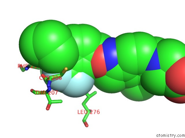
Mono view
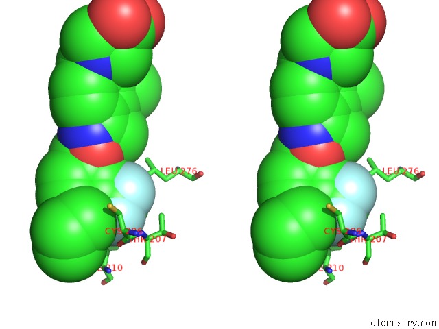
Stereo pair view
|
A full contact list of Fluorine with other atoms in the F binding
site number 2 of Cryo-Em Structure of Siponimod -Bound Sphingosine-1-Phosphate Receptor 1 in Complex with Gi Protein within 5.0Å range:
probe
|
atom
|
residue
|
distance (Å)
|
B
|
Occ
|
D:F401
b:124.3
occ:1.00
|
F27
|
D:J8C401
|
0.0
|
124.3
|
1.0
|
C25
|
D:J8C401
|
1.4
|
124.3
|
1.0
|
F26
|
D:J8C401
|
2.2
|
124.3
|
1.0
|
F28
|
D:J8C401
|
2.2
|
124.3
|
1.0
|
C15
|
D:J8C401
|
2.4
|
124.3
|
1.0
|
C19
|
D:J8C401
|
2.7
|
124.3
|
1.0
|
C16
|
D:J8C401
|
2.8
|
124.3
|
1.0
|
O
|
D:CYS206
|
3.2
|
124.1
|
1.0
|
C
|
D:CYS206
|
3.3
|
124.1
|
1.0
|
C20
|
D:J8C401
|
3.3
|
124.3
|
1.0
|
C14
|
D:J8C401
|
3.6
|
124.3
|
1.0
|
N
|
D:THR207
|
3.6
|
122.5
|
1.0
|
CB
|
D:CYS206
|
3.6
|
124.1
|
1.0
|
CA
|
D:THR207
|
3.8
|
122.5
|
1.0
|
CB
|
D:PHE210
|
3.9
|
110.7
|
1.0
|
C24
|
D:J8C401
|
4.0
|
124.3
|
1.0
|
CA
|
D:CYS206
|
4.0
|
124.1
|
1.0
|
CD2
|
D:LEU276
|
4.1
|
126.4
|
1.0
|
C17
|
D:J8C401
|
4.2
|
124.3
|
1.0
|
CG2
|
D:THR207
|
4.3
|
122.5
|
1.0
|
CG
|
D:PHE210
|
4.5
|
110.7
|
1.0
|
C21
|
D:J8C401
|
4.7
|
124.3
|
1.0
|
CB
|
D:THR207
|
4.7
|
122.5
|
1.0
|
C13
|
D:J8C401
|
4.7
|
124.3
|
1.0
|
C23
|
D:J8C401
|
4.8
|
124.3
|
1.0
|
C
|
D:THR207
|
4.9
|
122.5
|
1.0
|
C22
|
D:J8C401
|
4.9
|
124.3
|
1.0
|
CD2
|
D:PHE210
|
5.0
|
110.7
|
1.0
|
C18
|
D:J8C401
|
5.0
|
124.3
|
1.0
|
|
Fluorine binding site 3 out
of 3 in 7evy
Go back to
Fluorine Binding Sites List in 7evy
Fluorine binding site 3 out
of 3 in the Cryo-Em Structure of Siponimod -Bound Sphingosine-1-Phosphate Receptor 1 in Complex with Gi Protein
Mono view
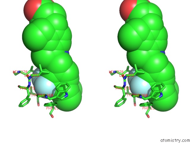
Stereo pair view
|
A full contact list of Fluorine with other atoms in the F binding
site number 3 of Cryo-Em Structure of Siponimod -Bound Sphingosine-1-Phosphate Receptor 1 in Complex with Gi Protein within 5.0Å range:
probe
|
atom
|
residue
|
distance (Å)
|
B
|
Occ
|
D:F401
b:124.3
occ:1.00
|
F28
|
D:J8C401
|
0.0
|
124.3
|
1.0
|
C25
|
D:J8C401
|
1.4
|
124.3
|
1.0
|
F26
|
D:J8C401
|
2.2
|
124.3
|
1.0
|
F27
|
D:J8C401
|
2.2
|
124.3
|
1.0
|
C15
|
D:J8C401
|
2.3
|
124.3
|
1.0
|
C14
|
D:J8C401
|
3.1
|
124.3
|
1.0
|
C16
|
D:J8C401
|
3.3
|
124.3
|
1.0
|
C19
|
D:J8C401
|
3.6
|
124.3
|
1.0
|
CD2
|
D:LEU276
|
4.1
|
126.4
|
1.0
|
CG
|
D:PHE210
|
4.2
|
110.7
|
1.0
|
CD2
|
D:PHE210
|
4.3
|
110.7
|
1.0
|
C13
|
D:J8C401
|
4.3
|
124.3
|
1.0
|
CB
|
D:PHE210
|
4.3
|
110.7
|
1.0
|
CD2
|
D:LEU272
|
4.4
|
120.6
|
1.0
|
C24
|
D:J8C401
|
4.4
|
124.3
|
1.0
|
C17
|
D:J8C401
|
4.4
|
124.3
|
1.0
|
CZ3
|
D:TRP269
|
4.5
|
107.7
|
1.0
|
CG
|
D:LEU272
|
4.6
|
120.6
|
1.0
|
CE3
|
D:TRP269
|
4.6
|
107.7
|
1.0
|
CD1
|
D:PHE210
|
4.6
|
110.7
|
1.0
|
CA
|
D:PHE273
|
4.8
|
120.3
|
1.0
|
CE2
|
D:PHE210
|
4.8
|
110.7
|
1.0
|
C20
|
D:J8C401
|
4.9
|
124.3
|
1.0
|
CD1
|
D:PHE273
|
4.9
|
120.3
|
1.0
|
C18
|
D:J8C401
|
4.9
|
124.3
|
1.0
|
CG2
|
D:THR207
|
4.9
|
122.5
|
1.0
|
N
|
D:PHE273
|
4.9
|
120.3
|
1.0
|
CB
|
D:PHE273
|
5.0
|
120.3
|
1.0
|
CA
|
D:THR207
|
5.0
|
122.5
|
1.0
|
|
Reference:
Y.Yuan,
G.Jia,
C.Wu,
W.Wang,
L.Cheng,
Q.Li,
Z.Li,
K.Luo,
S.Yang,
W.Yan,
Z.Su,
Z.Shao.
Structures of Signaling Complexes of Lipid Receptors S1PR1 and S1PR5 Reveal Mechanisms of Activation and Drug Recognition. Cell Res. 2021.
ISSN: ISSN 1001-0602
PubMed: 34526663
DOI: 10.1038/S41422-021-00566-X
Page generated: Fri Aug 2 06:46:39 2024
|