Fluorine »
PDB 7gba-7gkf »
7gch »
Fluorine in PDB 7gch: Structure of Chymotrypsin-*Trifluoromethyl Ketone Inhibitor Complexes. Comparison of Slowly and Rapidly Equilibrating Inhibitors
Enzymatic activity of Structure of Chymotrypsin-*Trifluoromethyl Ketone Inhibitor Complexes. Comparison of Slowly and Rapidly Equilibrating Inhibitors
All present enzymatic activity of Structure of Chymotrypsin-*Trifluoromethyl Ketone Inhibitor Complexes. Comparison of Slowly and Rapidly Equilibrating Inhibitors:
3.4.21.1;
3.4.21.1;
Protein crystallography data
The structure of Structure of Chymotrypsin-*Trifluoromethyl Ketone Inhibitor Complexes. Comparison of Slowly and Rapidly Equilibrating Inhibitors, PDB code: 7gch
was solved by
K.Brady,
D.Ringe,
R.H.Abeles,
with X-Ray Crystallography technique. A brief refinement statistics is given in the table below:
| Resolution Low / High (Å) | 10.00 / 1.80 |
| Space group | P 42 21 2 |
| Cell size a, b, c (Å), α, β, γ (°) | 69.300, 69.300, 97.600, 90.00, 90.00, 90.00 |
| R / Rfree (%) | n/a / n/a |
Fluorine Binding Sites:
The binding sites of Fluorine atom in the Structure of Chymotrypsin-*Trifluoromethyl Ketone Inhibitor Complexes. Comparison of Slowly and Rapidly Equilibrating Inhibitors
(pdb code 7gch). This binding sites where shown within
5.0 Angstroms radius around Fluorine atom.
In total 3 binding sites of Fluorine where determined in the Structure of Chymotrypsin-*Trifluoromethyl Ketone Inhibitor Complexes. Comparison of Slowly and Rapidly Equilibrating Inhibitors, PDB code: 7gch:
Jump to Fluorine binding site number: 1; 2; 3;
In total 3 binding sites of Fluorine where determined in the Structure of Chymotrypsin-*Trifluoromethyl Ketone Inhibitor Complexes. Comparison of Slowly and Rapidly Equilibrating Inhibitors, PDB code: 7gch:
Jump to Fluorine binding site number: 1; 2; 3;
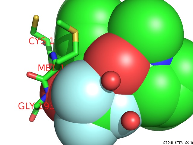
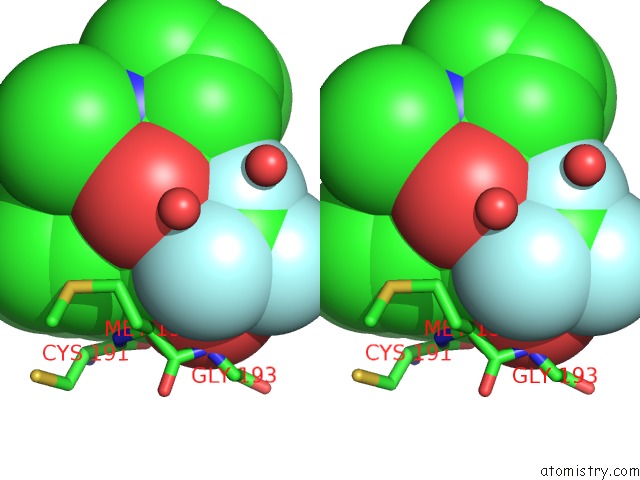
Fluorine binding site 1 out of 3 in 7gch
Go back to
Fluorine binding site 1 out
of 3 in the Structure of Chymotrypsin-*Trifluoromethyl Ketone Inhibitor Complexes. Comparison of Slowly and Rapidly Equilibrating Inhibitors
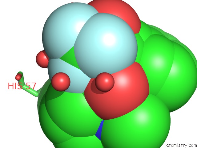
Mono view
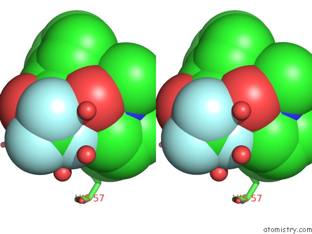
Stereo pair view
Mono view
Stereo pair view
A full contact list of Fluorine with other atoms in the F binding
site number 1 of Structure of Chymotrypsin-*Trifluoromethyl Ketone Inhibitor Complexes. Comparison of Slowly and Rapidly Equilibrating Inhibitors within 5.0Å range:
|
Fluorine binding site 2 out of 3 in 7gch
Go back to
Fluorine binding site 2 out
of 3 in the Structure of Chymotrypsin-*Trifluoromethyl Ketone Inhibitor Complexes. Comparison of Slowly and Rapidly Equilibrating Inhibitors
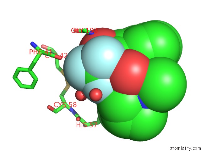
Mono view
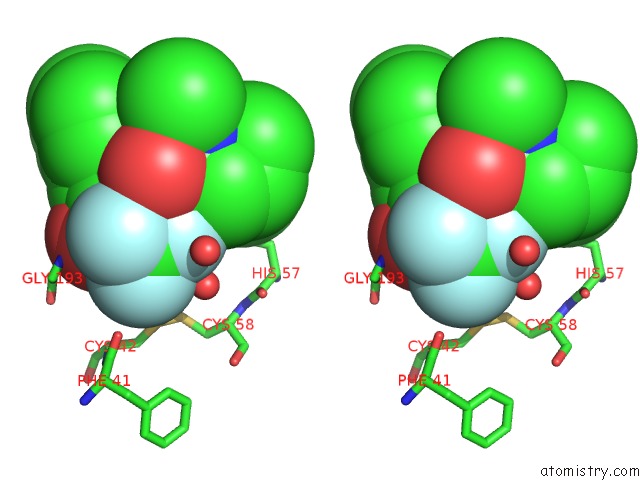
Stereo pair view
Mono view
Stereo pair view
A full contact list of Fluorine with other atoms in the F binding
site number 2 of Structure of Chymotrypsin-*Trifluoromethyl Ketone Inhibitor Complexes. Comparison of Slowly and Rapidly Equilibrating Inhibitors within 5.0Å range:
|
Fluorine binding site 3 out of 3 in 7gch
Go back to
Fluorine binding site 3 out
of 3 in the Structure of Chymotrypsin-*Trifluoromethyl Ketone Inhibitor Complexes. Comparison of Slowly and Rapidly Equilibrating Inhibitors
Mono view
Stereo pair view
Mono view
Stereo pair view
A full contact list of Fluorine with other atoms in the F binding
site number 3 of Structure of Chymotrypsin-*Trifluoromethyl Ketone Inhibitor Complexes. Comparison of Slowly and Rapidly Equilibrating Inhibitors within 5.0Å range:
|
Reference:
K.Brady,
A.Z.Wei,
D.Ringe,
R.H.Abeles.
Structure of Chymotrypsin-Trifluoromethyl Ketone Inhibitor Complexes: Comparison of Slowly and Rapidly Equilibrating Inhibitors. Biochemistry V. 29 7600 1990.
ISSN: ISSN 0006-2960
PubMed: 2271520
DOI: 10.1021/BI00485A009
Page generated: Tue Jul 15 20:10:14 2025
ISSN: ISSN 0006-2960
PubMed: 2271520
DOI: 10.1021/BI00485A009
Last articles
Fe in 2YXOFe in 2YRS
Fe in 2YXC
Fe in 2YNM
Fe in 2YVJ
Fe in 2YP1
Fe in 2YU2
Fe in 2YU1
Fe in 2YQB
Fe in 2YOO