Fluorine in PDB 7gro: Crystal Structure of Sars-Cov-2 Main Protease in Complex with Cpd-11
Enzymatic activity of Crystal Structure of Sars-Cov-2 Main Protease in Complex with Cpd-11
All present enzymatic activity of Crystal Structure of Sars-Cov-2 Main Protease in Complex with Cpd-11:
3.4.22.69;
3.4.22.69;
Protein crystallography data
The structure of Crystal Structure of Sars-Cov-2 Main Protease in Complex with Cpd-11, PDB code: 7gro
was solved by
C.-Y.Huang,
A.Metz,
M.Sharpe,
A.Sweeney,
with X-Ray Crystallography technique. A brief refinement statistics is given in the table below:
| Resolution Low / High (Å) | 56.68 / 1.55 |
| Space group | P 21 21 21 |
| Cell size a, b, c (Å), α, β, γ (°) | 67.62, 99.49, 103.93, 90, 90, 90 |
| R / Rfree (%) | 19.2 / 23.2 |
Other elements in 7gro:
The structure of Crystal Structure of Sars-Cov-2 Main Protease in Complex with Cpd-11 also contains other interesting chemical elements:
| Chlorine | (Cl) | 2 atoms |
| Sodium | (Na) | 2 atoms |
Fluorine Binding Sites:
The binding sites of Fluorine atom in the Crystal Structure of Sars-Cov-2 Main Protease in Complex with Cpd-11
(pdb code 7gro). This binding sites where shown within
5.0 Angstroms radius around Fluorine atom.
In total 3 binding sites of Fluorine where determined in the Crystal Structure of Sars-Cov-2 Main Protease in Complex with Cpd-11, PDB code: 7gro:
Jump to Fluorine binding site number: 1; 2; 3;
In total 3 binding sites of Fluorine where determined in the Crystal Structure of Sars-Cov-2 Main Protease in Complex with Cpd-11, PDB code: 7gro:
Jump to Fluorine binding site number: 1; 2; 3;
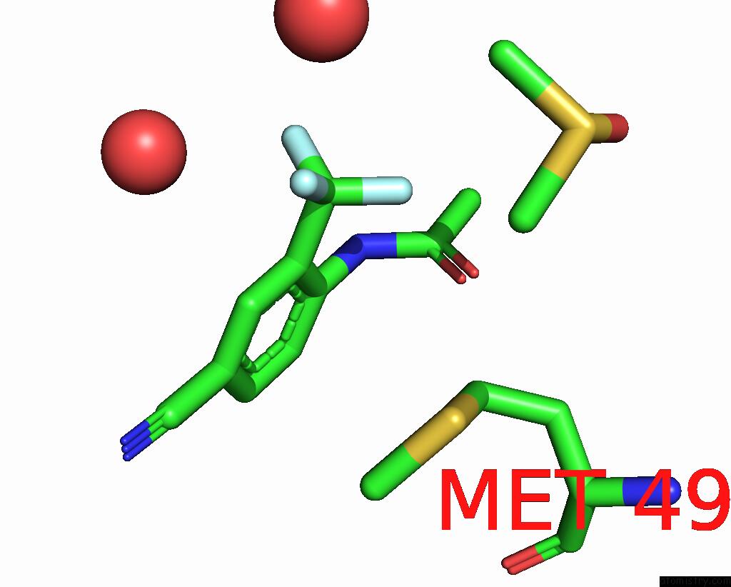
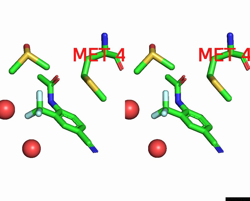
Fluorine binding site 1 out of 3 in 7gro
Go back to
Fluorine binding site 1 out
of 3 in the Crystal Structure of Sars-Cov-2 Main Protease in Complex with Cpd-11
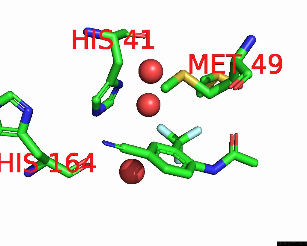
Mono view
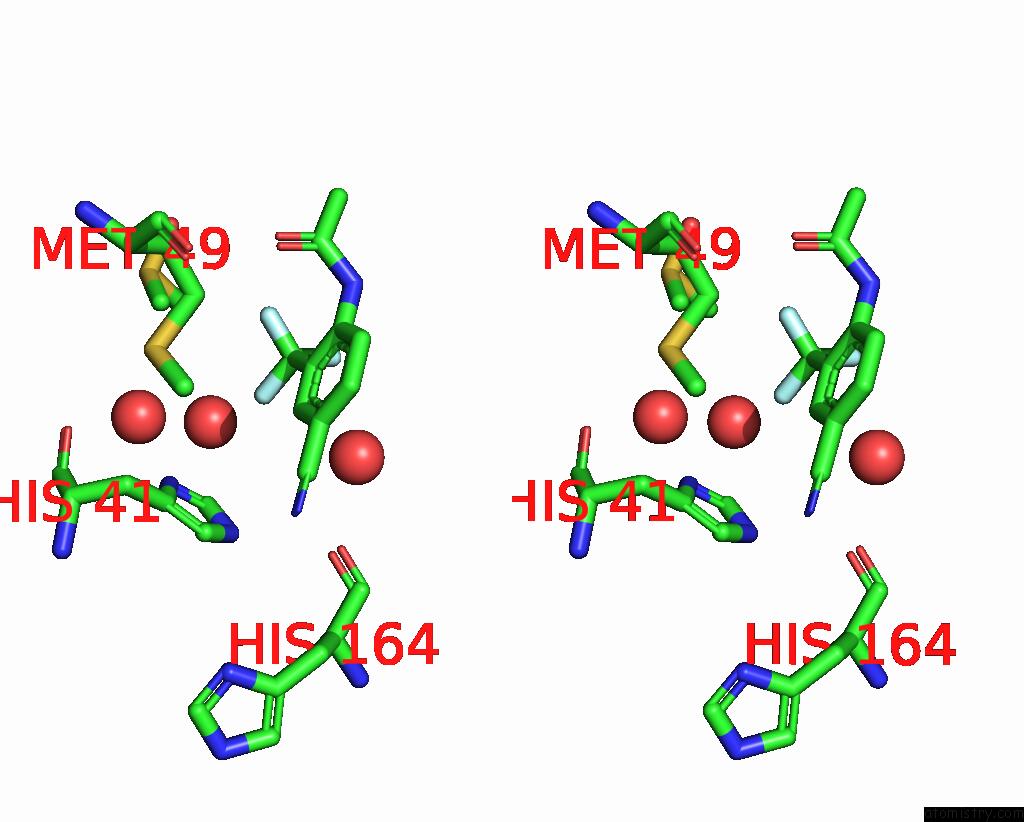
Stereo pair view
Mono view
Stereo pair view
A full contact list of Fluorine with other atoms in the F binding
site number 1 of Crystal Structure of Sars-Cov-2 Main Protease in Complex with Cpd-11 within 5.0Å range:
|
Fluorine binding site 2 out of 3 in 7gro
Go back to
Fluorine binding site 2 out
of 3 in the Crystal Structure of Sars-Cov-2 Main Protease in Complex with Cpd-11
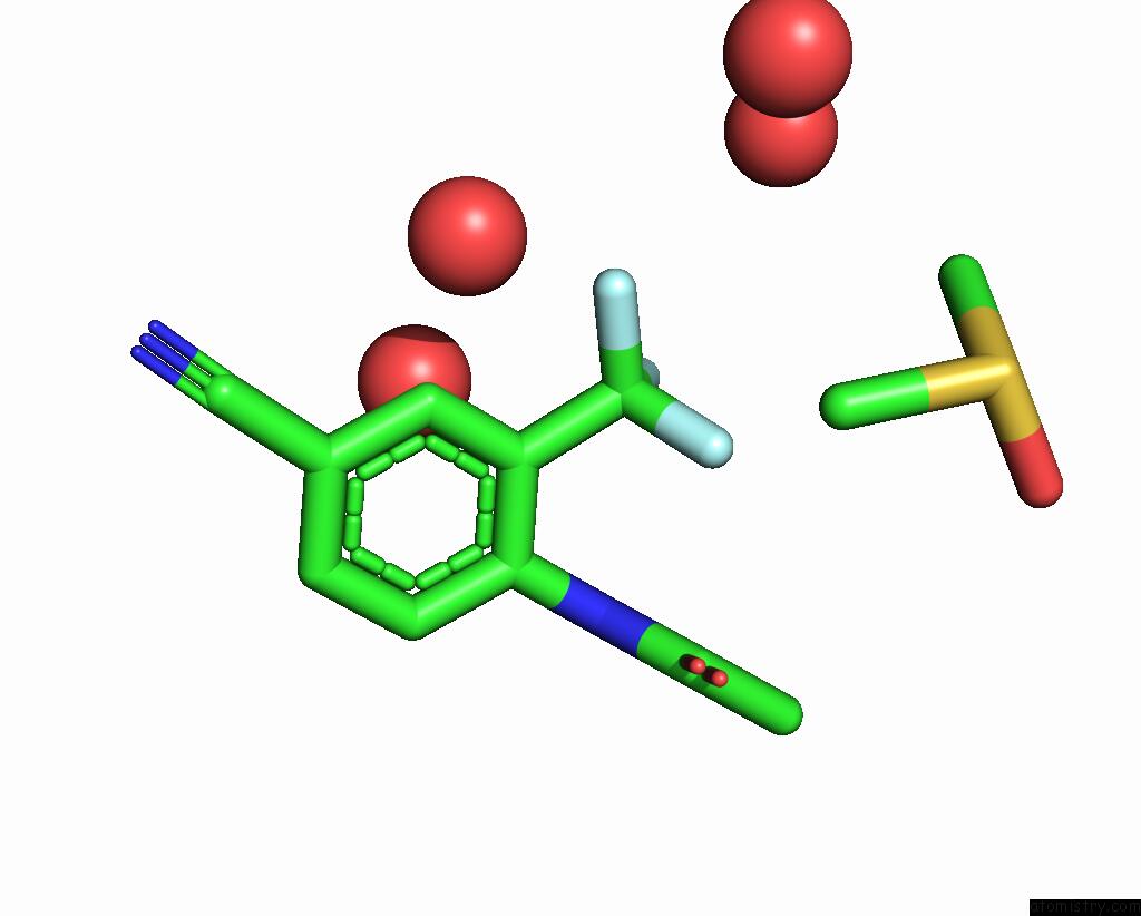
Mono view
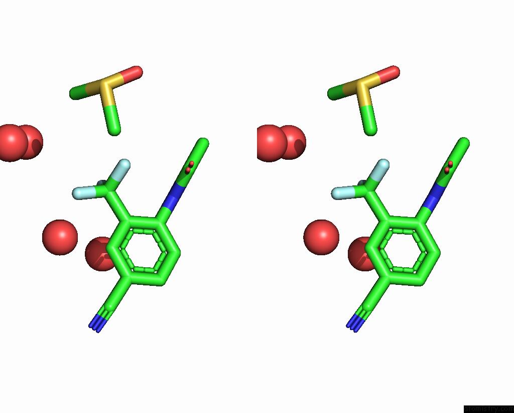
Stereo pair view
Mono view
Stereo pair view
A full contact list of Fluorine with other atoms in the F binding
site number 2 of Crystal Structure of Sars-Cov-2 Main Protease in Complex with Cpd-11 within 5.0Å range:
|
Fluorine binding site 3 out of 3 in 7gro
Go back to
Fluorine binding site 3 out
of 3 in the Crystal Structure of Sars-Cov-2 Main Protease in Complex with Cpd-11
Mono view
Stereo pair view
Mono view
Stereo pair view
A full contact list of Fluorine with other atoms in the F binding
site number 3 of Crystal Structure of Sars-Cov-2 Main Protease in Complex with Cpd-11 within 5.0Å range:
|
Reference:
C.Y.Huang,
A.Metz,
R.Lange,
N.Artico,
C.Potot,
J.Hazemann,
M.Muller,
M.Dos Santos,
A.Chambovey,
D.Ritz,
D.Eris,
S.Meyer,
G.Bourquin,
M.Sharpe,
A.Mac Sweeney.
Fragment-Based Screening Targeting An Open Form of the Sars-Cov-2 Main Protease Binding Pocket. Acta Crystallogr D Struct V. 80 123 2024BIOL.
ISSN: ISSN 2059-7983
PubMed: 38289714
DOI: 10.1107/S2059798324000329
Page generated: Fri Aug 2 07:45:05 2024
ISSN: ISSN 2059-7983
PubMed: 38289714
DOI: 10.1107/S2059798324000329
Last articles
Zn in 9MJ5Zn in 9HNW
Zn in 9G0L
Zn in 9FNE
Zn in 9DZN
Zn in 9E0I
Zn in 9D32
Zn in 9DAK
Zn in 8ZXC
Zn in 8ZUF