Fluorine »
PDB 7ho4-7jx9 »
7hog »
Fluorine in PDB 7hog: Group Deposition For Crystallographic Fragment Screening of Coxsackievirus A16 (G-10) 2A Protease -- Crystal Structure of Coxsackievirus A16 (G-10) 2A Protease in Complex with Z1216861874 (A71EV2A-X0525)
Enzymatic activity of Group Deposition For Crystallographic Fragment Screening of Coxsackievirus A16 (G-10) 2A Protease -- Crystal Structure of Coxsackievirus A16 (G-10) 2A Protease in Complex with Z1216861874 (A71EV2A-X0525)
All present enzymatic activity of Group Deposition For Crystallographic Fragment Screening of Coxsackievirus A16 (G-10) 2A Protease -- Crystal Structure of Coxsackievirus A16 (G-10) 2A Protease in Complex with Z1216861874 (A71EV2A-X0525):
3.4.22.29;
3.4.22.29;
Protein crystallography data
The structure of Group Deposition For Crystallographic Fragment Screening of Coxsackievirus A16 (G-10) 2A Protease -- Crystal Structure of Coxsackievirus A16 (G-10) 2A Protease in Complex with Z1216861874 (A71EV2A-X0525), PDB code: 7hog
was solved by
R.M.Lithgo,
M.Fairhead,
L.Koekemoer,
B.H.Balcomb,
E.Capkin,
A.V.Chandran,
M.Golding,
A.S.Godoy,
J.C.Aschenbrenner,
P.G.Marples,
X.Ni,
W.Thompson,
C.W.E.Tomlinson,
C.Wild,
M.Winokan,
M.-A.E.Xavier,
D.Fearon,
F.Von Delft,
with X-Ray Crystallography technique. A brief refinement statistics is given in the table below:
| Resolution Low / High (Å) | 47.20 / 1.19 |
| Space group | C 1 2 1 |
| Cell size a, b, c (Å), α, β, γ (°) | 86.088, 56.539, 32.421, 90, 95.26, 90 |
| R / Rfree (%) | 17 / 18.7 |
Other elements in 7hog:
The structure of Group Deposition For Crystallographic Fragment Screening of Coxsackievirus A16 (G-10) 2A Protease -- Crystal Structure of Coxsackievirus A16 (G-10) 2A Protease in Complex with Z1216861874 (A71EV2A-X0525) also contains other interesting chemical elements:
| Zinc | (Zn) | 1 atom |
Fluorine Binding Sites:
The binding sites of Fluorine atom in the Group Deposition For Crystallographic Fragment Screening of Coxsackievirus A16 (G-10) 2A Protease -- Crystal Structure of Coxsackievirus A16 (G-10) 2A Protease in Complex with Z1216861874 (A71EV2A-X0525)
(pdb code 7hog). This binding sites where shown within
5.0 Angstroms radius around Fluorine atom.
In total 2 binding sites of Fluorine where determined in the Group Deposition For Crystallographic Fragment Screening of Coxsackievirus A16 (G-10) 2A Protease -- Crystal Structure of Coxsackievirus A16 (G-10) 2A Protease in Complex with Z1216861874 (A71EV2A-X0525), PDB code: 7hog:
Jump to Fluorine binding site number: 1; 2;
Fluorine binding site 1 out of 2 in 7hog
Go back to
Fluorine binding site 1 out
of 2 in the Group Deposition For Crystallographic Fragment Screening of Coxsackievirus A16 (G-10) 2A Protease -- Crystal Structure of Coxsackievirus A16 (G-10) 2A Protease in Complex with Z1216861874 (A71EV2A-X0525)
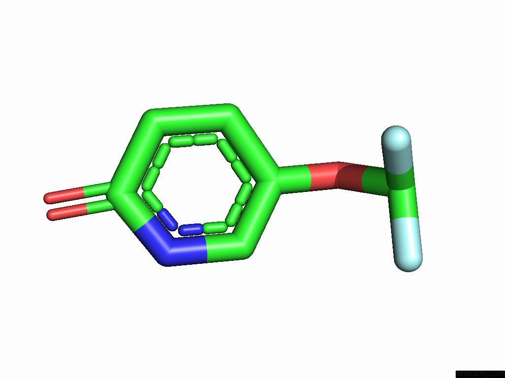
Mono view
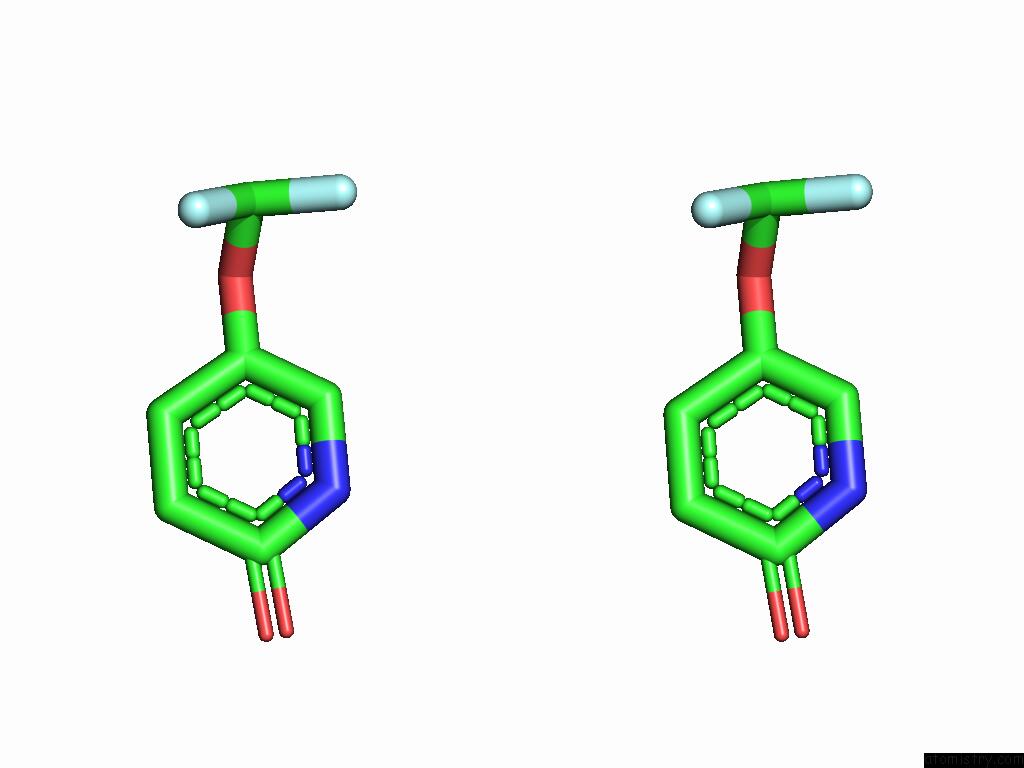
Stereo pair view
Mono view
Stereo pair view
A full contact list of Fluorine with other atoms in the F binding
site number 1 of Group Deposition For Crystallographic Fragment Screening of Coxsackievirus A16 (G-10) 2A Protease -- Crystal Structure of Coxsackievirus A16 (G-10) 2A Protease in Complex with Z1216861874 (A71EV2A-X0525) within 5.0Å range:
|
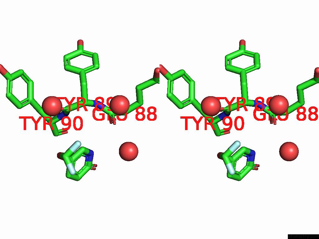
Fluorine binding site 2 out of 2 in 7hog
Go back to
Fluorine binding site 2 out
of 2 in the Group Deposition For Crystallographic Fragment Screening of Coxsackievirus A16 (G-10) 2A Protease -- Crystal Structure of Coxsackievirus A16 (G-10) 2A Protease in Complex with Z1216861874 (A71EV2A-X0525)
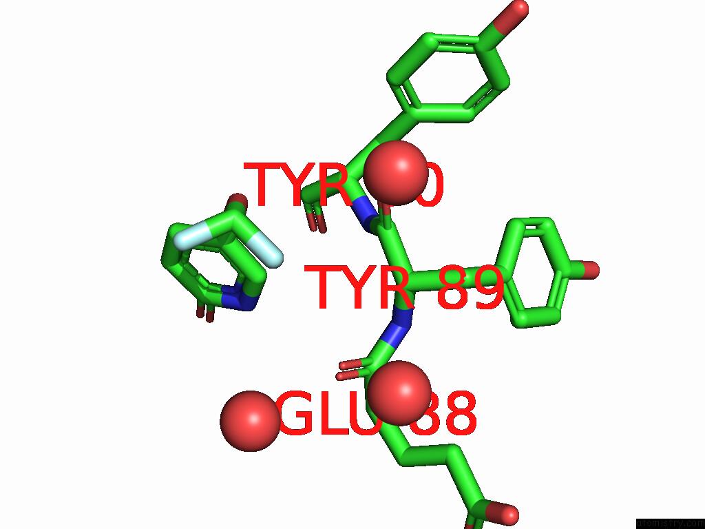
Mono view
Stereo pair view
Mono view
Stereo pair view
A full contact list of Fluorine with other atoms in the F binding
site number 2 of Group Deposition For Crystallographic Fragment Screening of Coxsackievirus A16 (G-10) 2A Protease -- Crystal Structure of Coxsackievirus A16 (G-10) 2A Protease in Complex with Z1216861874 (A71EV2A-X0525) within 5.0Å range:
|
Reference:
R.M.Lithgo,
M.Fairhead,
L.Koekemoer,
B.H.Balcomb,
E.Capkin,
A.V.Chandran,
M.Golding,
A.S.Godoy,
J.C.Aschenbrenner,
P.G.Marples,
X.Ni,
W.Thompson,
C.W.E.Tomlinson,
C.Wild,
M.Winokan,
M.-A.E.Xavier,
D.Fearon,
F.Von Delft.
Group Deposition For Crystallographic Fragment Screening of Coxsackievirus A16 (G-10) 2A Protease To Be Published.
Page generated: Sat Feb 8 18:12:04 2025
Last articles
As in 9E9GAl in 9B2D
Al in 9ED3
Zn in 9IRQ
Zn in 9IYX
Zn in 9J8P
Zn in 9IUU
Zn in 9GBF
Zn in 9G2V
Zn in 9G2L