Fluorine »
PDB 7kqd-7kzy »
7kvu »
Fluorine in PDB 7kvu: Crystal Structure of Squash Rna Aptamer in Complex with Dfhbi-1T
Protein crystallography data
The structure of Crystal Structure of Squash Rna Aptamer in Complex with Dfhbi-1T, PDB code: 7kvu
was solved by
L.Truong,
A.R.Ferre-D'amare,
with X-Ray Crystallography technique. A brief refinement statistics is given in the table below:
| Resolution Low / High (Å) | 46.46 / 2.68 |
| Space group | P 42 21 2 |
| Cell size a, b, c (Å), α, β, γ (°) | 103.731, 103.731, 51.959, 90, 90, 90 |
| R / Rfree (%) | 20.9 / 24.7 |
Other elements in 7kvu:
The structure of Crystal Structure of Squash Rna Aptamer in Complex with Dfhbi-1T also contains other interesting chemical elements:
| Potassium | (K) | 3 atoms |
| Magnesium | (Mg) | 4 atoms |
Fluorine Binding Sites:
The binding sites of Fluorine atom in the Crystal Structure of Squash Rna Aptamer in Complex with Dfhbi-1T
(pdb code 7kvu). This binding sites where shown within
5.0 Angstroms radius around Fluorine atom.
In total 5 binding sites of Fluorine where determined in the Crystal Structure of Squash Rna Aptamer in Complex with Dfhbi-1T, PDB code: 7kvu:
Jump to Fluorine binding site number: 1; 2; 3; 4; 5;
In total 5 binding sites of Fluorine where determined in the Crystal Structure of Squash Rna Aptamer in Complex with Dfhbi-1T, PDB code: 7kvu:
Jump to Fluorine binding site number: 1; 2; 3; 4; 5;
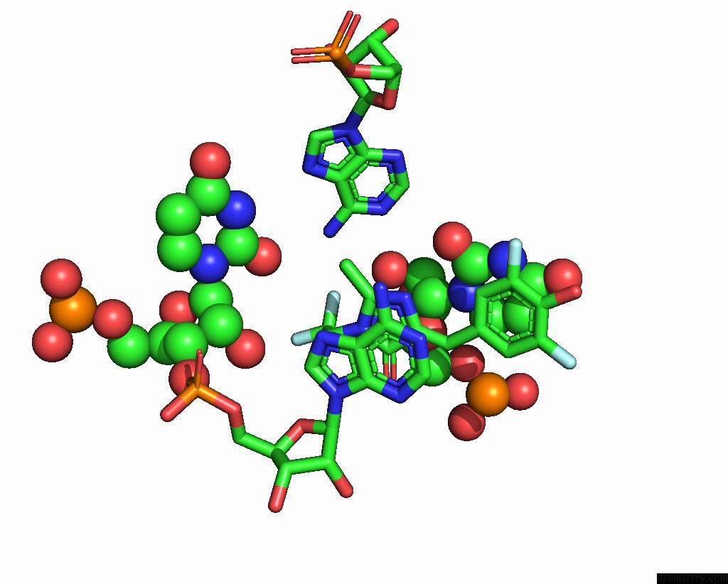
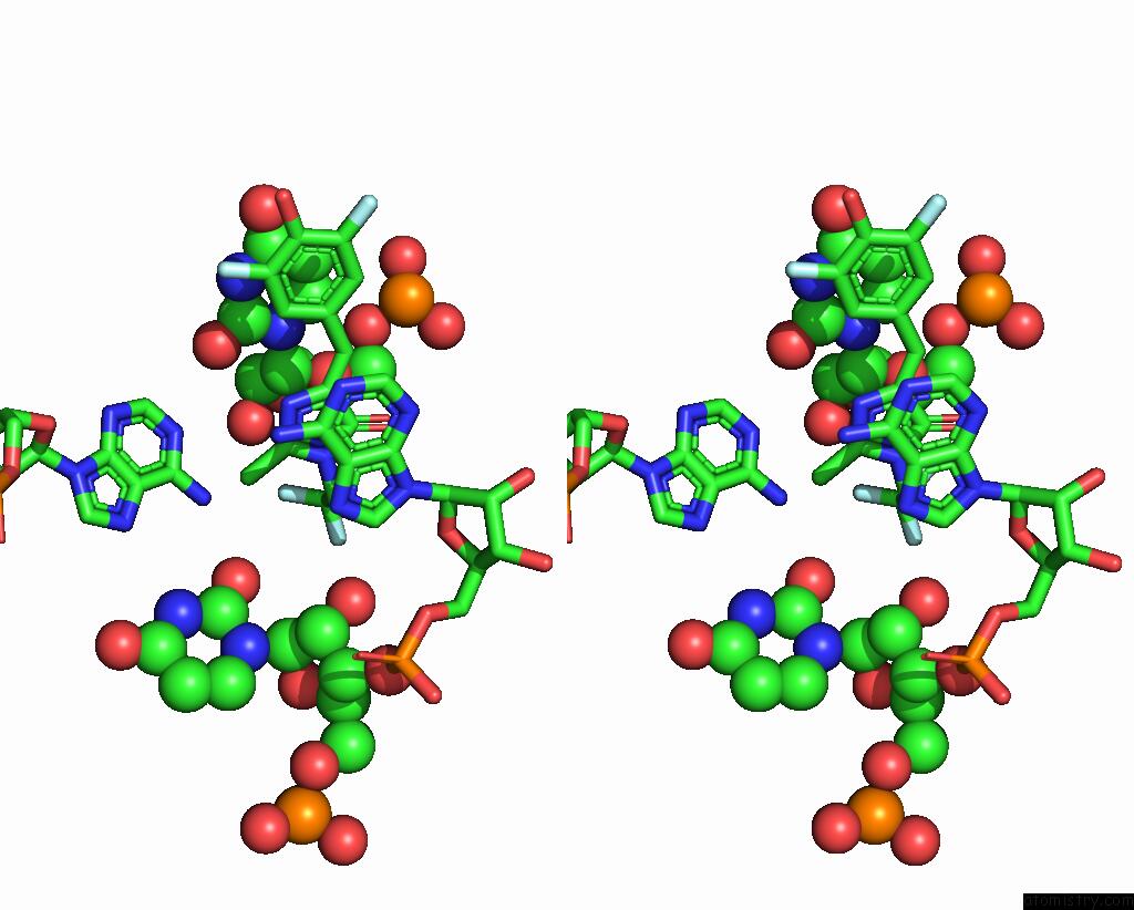
Fluorine binding site 1 out of 5 in 7kvu
Go back to
Fluorine binding site 1 out
of 5 in the Crystal Structure of Squash Rna Aptamer in Complex with Dfhbi-1T
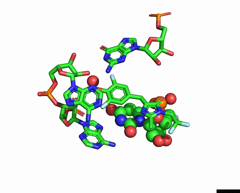
Mono view
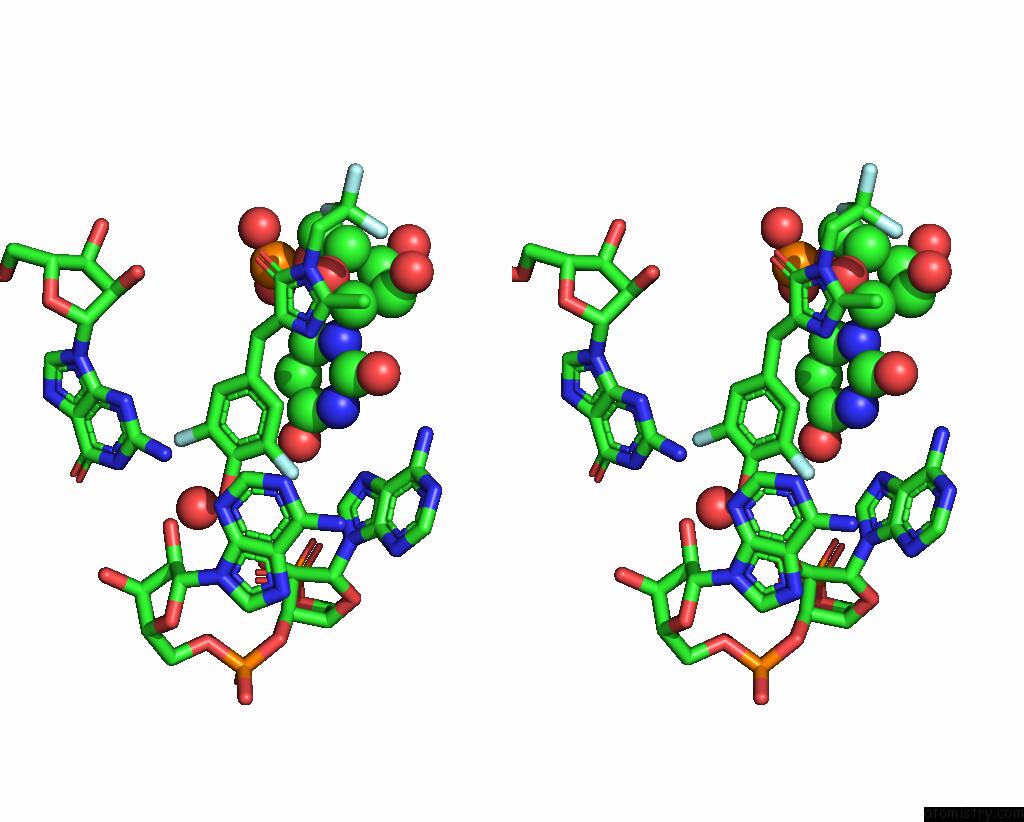
Stereo pair view
Mono view
Stereo pair view
A full contact list of Fluorine with other atoms in the F binding
site number 1 of Crystal Structure of Squash Rna Aptamer in Complex with Dfhbi-1T within 5.0Å range:
|
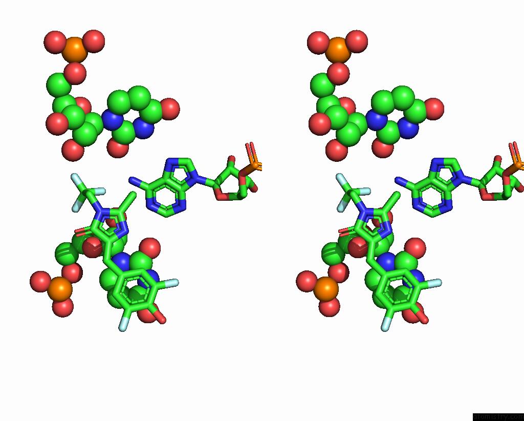
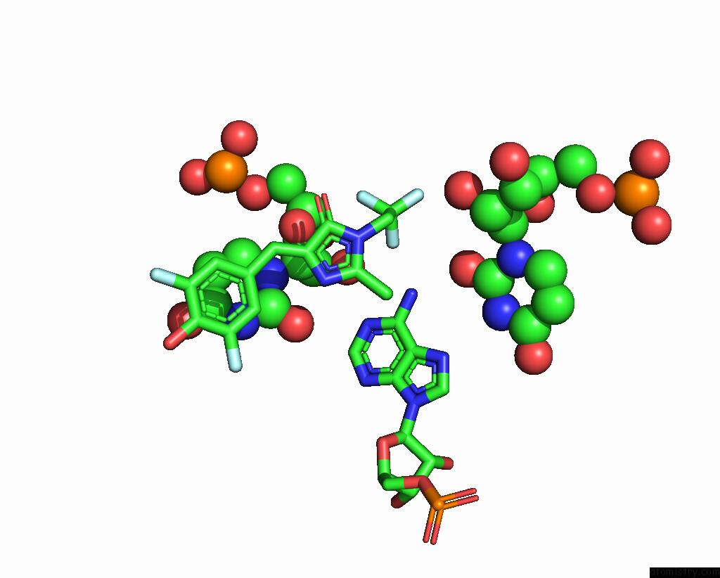
Fluorine binding site 2 out of 5 in 7kvu
Go back to
Fluorine binding site 2 out
of 5 in the Crystal Structure of Squash Rna Aptamer in Complex with Dfhbi-1T
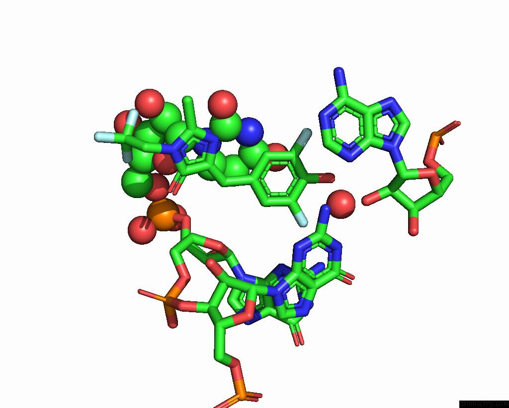
Mono view

Stereo pair view
Mono view
Stereo pair view
A full contact list of Fluorine with other atoms in the F binding
site number 2 of Crystal Structure of Squash Rna Aptamer in Complex with Dfhbi-1T within 5.0Å range:
|
Fluorine binding site 3 out of 5 in 7kvu
Go back to
Fluorine binding site 3 out
of 5 in the Crystal Structure of Squash Rna Aptamer in Complex with Dfhbi-1T

Mono view
Stereo pair view
Mono view
Stereo pair view
A full contact list of Fluorine with other atoms in the F binding
site number 3 of Crystal Structure of Squash Rna Aptamer in Complex with Dfhbi-1T within 5.0Å range:
|
Fluorine binding site 4 out of 5 in 7kvu
Go back to
Fluorine binding site 4 out
of 5 in the Crystal Structure of Squash Rna Aptamer in Complex with Dfhbi-1T
Mono view
Stereo pair view
Mono view
Stereo pair view
A full contact list of Fluorine with other atoms in the F binding
site number 4 of Crystal Structure of Squash Rna Aptamer in Complex with Dfhbi-1T within 5.0Å range:
|
Fluorine binding site 5 out of 5 in 7kvu
Go back to
Fluorine binding site 5 out
of 5 in the Crystal Structure of Squash Rna Aptamer in Complex with Dfhbi-1T
Mono view

Stereo pair view
Mono view
Stereo pair view
A full contact list of Fluorine with other atoms in the F binding
site number 5 of Crystal Structure of Squash Rna Aptamer in Complex with Dfhbi-1T within 5.0Å range:
|
Reference:
L.Truong,
H.Kooshapur,
S.K.Dey,
X.Li,
N.Tjandra,
S.R.Jaffrey,
A.R.Ferre-D'amare.
The Fluorescent Aptamer Squash Extensively Repurposes the Adenine Riboswitch Fold. Nat.Chem.Biol. V. 18 191 2022.
ISSN: ESSN 1552-4469
PubMed: 34937911
DOI: 10.1038/S41589-021-00931-2
Page generated: Tue Jul 15 20:57:15 2025
ISSN: ESSN 1552-4469
PubMed: 34937911
DOI: 10.1038/S41589-021-00931-2
Last articles
Fe in 2YXOFe in 2YRS
Fe in 2YXC
Fe in 2YNM
Fe in 2YVJ
Fe in 2YP1
Fe in 2YU2
Fe in 2YU1
Fe in 2YQB
Fe in 2YOO