Fluorine in PDB 7nor: Crystal Structure of Mycobacterium Tuberculosis Argf in Complex with 2-Fluoro-4-Hydroxybenzonitrile.
Enzymatic activity of Crystal Structure of Mycobacterium Tuberculosis Argf in Complex with 2-Fluoro-4-Hydroxybenzonitrile.
All present enzymatic activity of Crystal Structure of Mycobacterium Tuberculosis Argf in Complex with 2-Fluoro-4-Hydroxybenzonitrile.:
2.1.3.3;
2.1.3.3;
Protein crystallography data
The structure of Crystal Structure of Mycobacterium Tuberculosis Argf in Complex with 2-Fluoro-4-Hydroxybenzonitrile., PDB code: 7nor
was solved by
V.Mendes,
P.Gupta,
A.Burgess,
V.Sebastian-Perez,
E.Cattermole,
C.Meghir,
T.L.Blundell,
with X-Ray Crystallography technique. A brief refinement statistics is given in the table below:
| Resolution Low / High (Å) | 46.08 / 1.59 |
| Space group | P 1 21 1 |
| Cell size a, b, c (Å), α, β, γ (°) | 91.453, 142.734, 97.697, 90, 117.5, 90 |
| R / Rfree (%) | 16.7 / 18.6 |
Fluorine Binding Sites:
The binding sites of Fluorine atom in the Crystal Structure of Mycobacterium Tuberculosis Argf in Complex with 2-Fluoro-4-Hydroxybenzonitrile.
(pdb code 7nor). This binding sites where shown within
5.0 Angstroms radius around Fluorine atom.
In total 4 binding sites of Fluorine where determined in the Crystal Structure of Mycobacterium Tuberculosis Argf in Complex with 2-Fluoro-4-Hydroxybenzonitrile., PDB code: 7nor:
Jump to Fluorine binding site number: 1; 2; 3; 4;
In total 4 binding sites of Fluorine where determined in the Crystal Structure of Mycobacterium Tuberculosis Argf in Complex with 2-Fluoro-4-Hydroxybenzonitrile., PDB code: 7nor:
Jump to Fluorine binding site number: 1; 2; 3; 4;
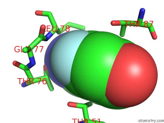
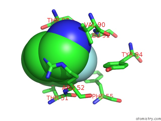
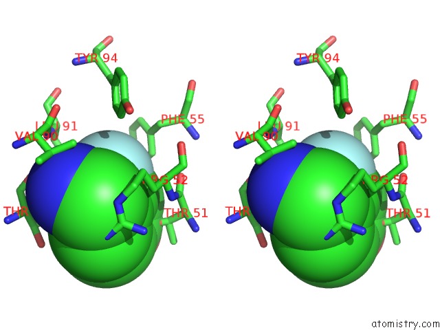
Fluorine binding site 1 out of 4 in 7nor
Go back to
Fluorine binding site 1 out
of 4 in the Crystal Structure of Mycobacterium Tuberculosis Argf in Complex with 2-Fluoro-4-Hydroxybenzonitrile.
Mono view
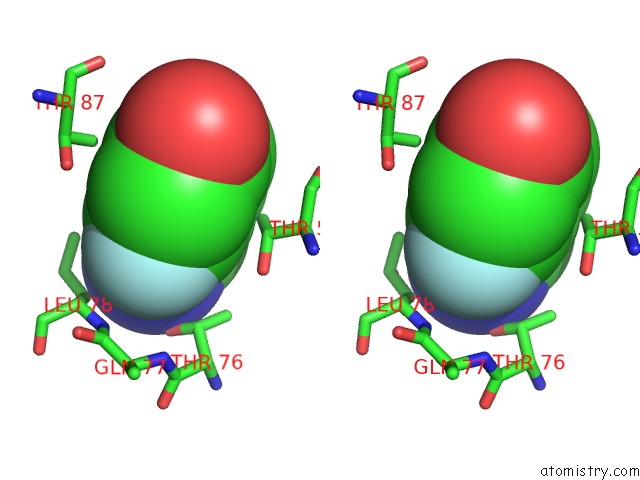
Stereo pair view
Mono view
Stereo pair view
A full contact list of Fluorine with other atoms in the F binding
site number 1 of Crystal Structure of Mycobacterium Tuberculosis Argf in Complex with 2-Fluoro-4-Hydroxybenzonitrile. within 5.0Å range:
|
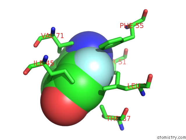
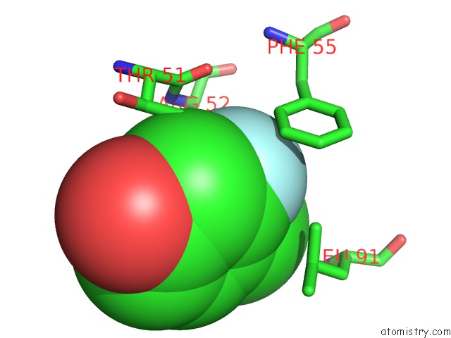
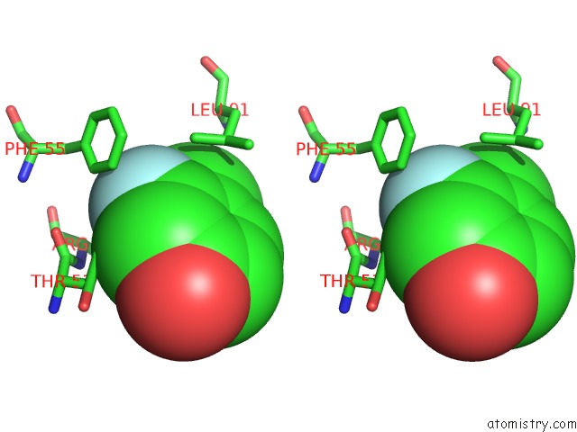
Fluorine binding site 2 out of 4 in 7nor
Go back to
Fluorine binding site 2 out
of 4 in the Crystal Structure of Mycobacterium Tuberculosis Argf in Complex with 2-Fluoro-4-Hydroxybenzonitrile.
Mono view
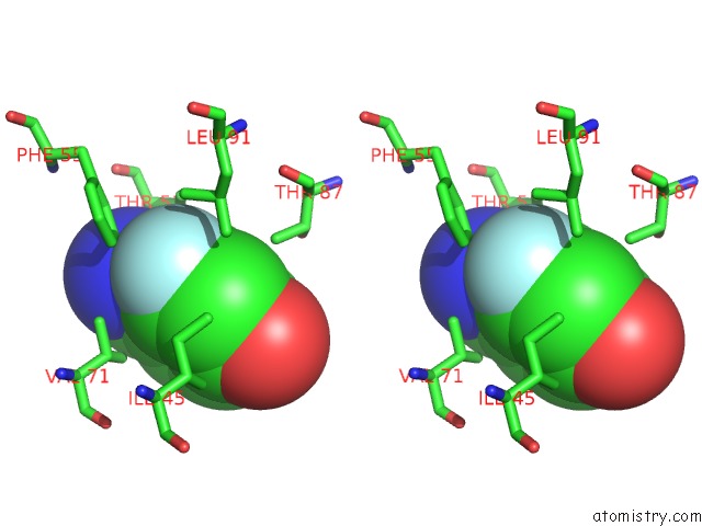
Stereo pair view
Mono view
Stereo pair view
A full contact list of Fluorine with other atoms in the F binding
site number 2 of Crystal Structure of Mycobacterium Tuberculosis Argf in Complex with 2-Fluoro-4-Hydroxybenzonitrile. within 5.0Å range:
|
Fluorine binding site 3 out of 4 in 7nor
Go back to
Fluorine binding site 3 out
of 4 in the Crystal Structure of Mycobacterium Tuberculosis Argf in Complex with 2-Fluoro-4-Hydroxybenzonitrile.
Mono view
Stereo pair view
Mono view
Stereo pair view
A full contact list of Fluorine with other atoms in the F binding
site number 3 of Crystal Structure of Mycobacterium Tuberculosis Argf in Complex with 2-Fluoro-4-Hydroxybenzonitrile. within 5.0Å range:
|
Fluorine binding site 4 out of 4 in 7nor
Go back to
Fluorine binding site 4 out
of 4 in the Crystal Structure of Mycobacterium Tuberculosis Argf in Complex with 2-Fluoro-4-Hydroxybenzonitrile.
Mono view
Stereo pair view
Mono view
Stereo pair view
A full contact list of Fluorine with other atoms in the F binding
site number 4 of Crystal Structure of Mycobacterium Tuberculosis Argf in Complex with 2-Fluoro-4-Hydroxybenzonitrile. within 5.0Å range:
|
Reference:
P.Gupta,
S.E.Thomas,
S.A.Zaidan,
M.A.Pasillas,
J.Cory-Wright,
V.Sebastian-Perez,
A.Burgess,
E.Cattermole,
C.Meghir,
C.Abell,
A.G.Coyne,
W.R.Jacobs,
T.L.Blundell,
S.Tiwari,
V.Mendes.
A Fragment-Based Approach to Assess the Ligandability of Argb, Argc, Argd and Argf in the L-Arginine Biosynthetic Pathway of Mycobacterium Tuberculosis Comput Struct Biotechnol J V. 19 3491 2021.
ISSN: ESSN 2001-0370
DOI: 10.1016/J.CSBJ.2021.06.006
Page generated: Fri Aug 2 10:12:16 2024
ISSN: ESSN 2001-0370
DOI: 10.1016/J.CSBJ.2021.06.006
Last articles
Zn in 9MJ5Zn in 9HNW
Zn in 9G0L
Zn in 9FNE
Zn in 9DZN
Zn in 9E0I
Zn in 9D32
Zn in 9DAK
Zn in 8ZXC
Zn in 8ZUF