Fluorine »
PDB 7ugj-7v3s »
7uju »
Fluorine in PDB 7uju: Room-Temperature X-Ray Structure of Monomeric Sars-Cov-2 Main Protease Catalytic Domain (MPRO1-196) in Complex with Nirmatrelvir
Enzymatic activity of Room-Temperature X-Ray Structure of Monomeric Sars-Cov-2 Main Protease Catalytic Domain (MPRO1-196) in Complex with Nirmatrelvir
All present enzymatic activity of Room-Temperature X-Ray Structure of Monomeric Sars-Cov-2 Main Protease Catalytic Domain (MPRO1-196) in Complex with Nirmatrelvir:
3.4.22.69;
3.4.22.69;
Protein crystallography data
The structure of Room-Temperature X-Ray Structure of Monomeric Sars-Cov-2 Main Protease Catalytic Domain (MPRO1-196) in Complex with Nirmatrelvir, PDB code: 7uju
was solved by
A.Kovalevsky,
D.W.Kneller,
L.Coates,
with X-Ray Crystallography technique. A brief refinement statistics is given in the table below:
| Resolution Low / High (Å) | 26.03 / 1.85 |
| Space group | P 1 21 1 |
| Cell size a, b, c (Å), α, β, γ (°) | 52.726, 62.605, 58.965, 90, 99.08, 90 |
| R / Rfree (%) | 15.4 / 17.5 |
Fluorine Binding Sites:
The binding sites of Fluorine atom in the Room-Temperature X-Ray Structure of Monomeric Sars-Cov-2 Main Protease Catalytic Domain (MPRO1-196) in Complex with Nirmatrelvir
(pdb code 7uju). This binding sites where shown within
5.0 Angstroms radius around Fluorine atom.
In total 6 binding sites of Fluorine where determined in the Room-Temperature X-Ray Structure of Monomeric Sars-Cov-2 Main Protease Catalytic Domain (MPRO1-196) in Complex with Nirmatrelvir, PDB code: 7uju:
Jump to Fluorine binding site number: 1; 2; 3; 4; 5; 6;
In total 6 binding sites of Fluorine where determined in the Room-Temperature X-Ray Structure of Monomeric Sars-Cov-2 Main Protease Catalytic Domain (MPRO1-196) in Complex with Nirmatrelvir, PDB code: 7uju:
Jump to Fluorine binding site number: 1; 2; 3; 4; 5; 6;
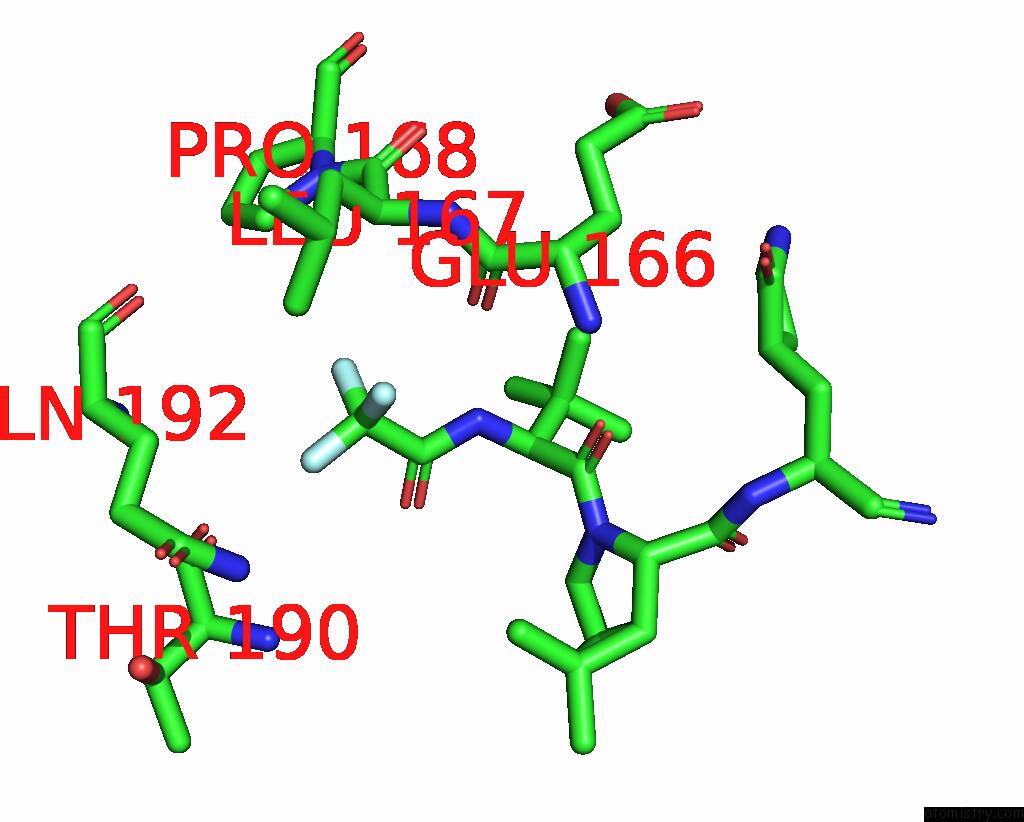
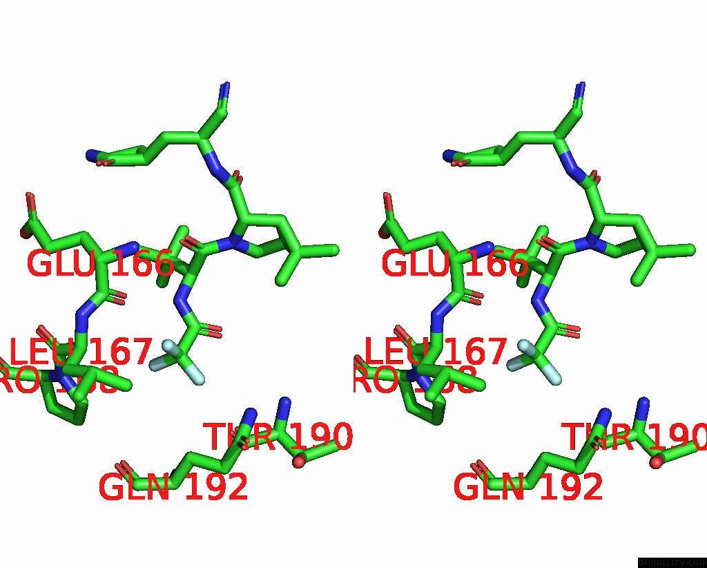
Fluorine binding site 1 out of 6 in 7uju
Go back to
Fluorine binding site 1 out
of 6 in the Room-Temperature X-Ray Structure of Monomeric Sars-Cov-2 Main Protease Catalytic Domain (MPRO1-196) in Complex with Nirmatrelvir
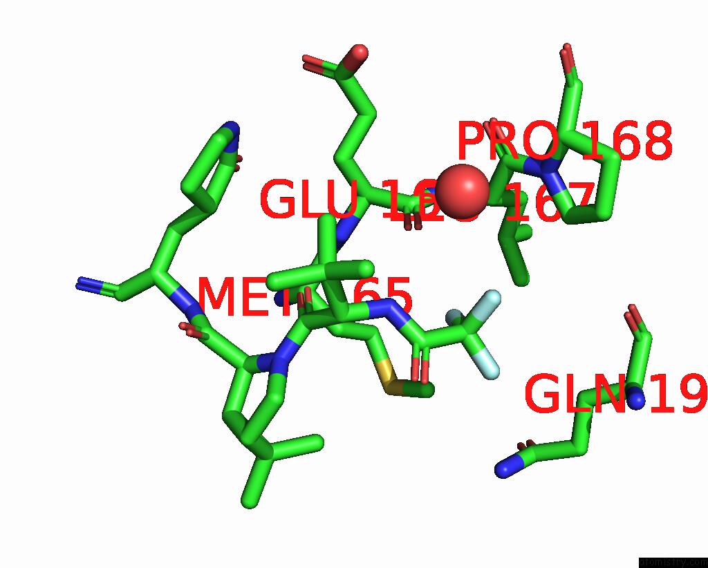
Mono view

Stereo pair view
Mono view
Stereo pair view
A full contact list of Fluorine with other atoms in the F binding
site number 1 of Room-Temperature X-Ray Structure of Monomeric Sars-Cov-2 Main Protease Catalytic Domain (MPRO1-196) in Complex with Nirmatrelvir within 5.0Å range:
|
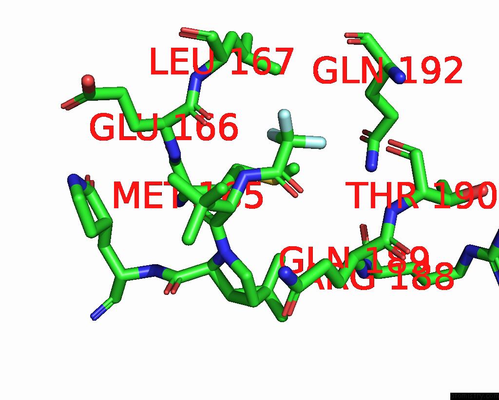
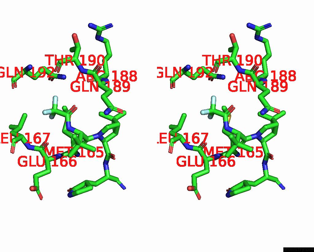
Fluorine binding site 2 out of 6 in 7uju
Go back to
Fluorine binding site 2 out
of 6 in the Room-Temperature X-Ray Structure of Monomeric Sars-Cov-2 Main Protease Catalytic Domain (MPRO1-196) in Complex with Nirmatrelvir

Mono view

Stereo pair view
Mono view
Stereo pair view
A full contact list of Fluorine with other atoms in the F binding
site number 2 of Room-Temperature X-Ray Structure of Monomeric Sars-Cov-2 Main Protease Catalytic Domain (MPRO1-196) in Complex with Nirmatrelvir within 5.0Å range:
|
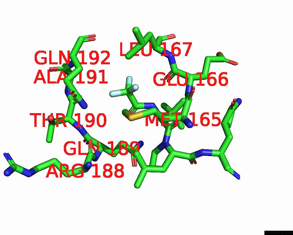
Fluorine binding site 3 out of 6 in 7uju
Go back to
Fluorine binding site 3 out
of 6 in the Room-Temperature X-Ray Structure of Monomeric Sars-Cov-2 Main Protease Catalytic Domain (MPRO1-196) in Complex with Nirmatrelvir
Mono view
Stereo pair view
Mono view
Stereo pair view
A full contact list of Fluorine with other atoms in the F binding
site number 3 of Room-Temperature X-Ray Structure of Monomeric Sars-Cov-2 Main Protease Catalytic Domain (MPRO1-196) in Complex with Nirmatrelvir within 5.0Å range:
|
Fluorine binding site 4 out of 6 in 7uju
Go back to
Fluorine binding site 4 out
of 6 in the Room-Temperature X-Ray Structure of Monomeric Sars-Cov-2 Main Protease Catalytic Domain (MPRO1-196) in Complex with Nirmatrelvir
Mono view

Stereo pair view
Mono view
Stereo pair view
A full contact list of Fluorine with other atoms in the F binding
site number 4 of Room-Temperature X-Ray Structure of Monomeric Sars-Cov-2 Main Protease Catalytic Domain (MPRO1-196) in Complex with Nirmatrelvir within 5.0Å range:
|
Fluorine binding site 5 out of 6 in 7uju
Go back to
Fluorine binding site 5 out
of 6 in the Room-Temperature X-Ray Structure of Monomeric Sars-Cov-2 Main Protease Catalytic Domain (MPRO1-196) in Complex with Nirmatrelvir
Mono view
Stereo pair view
Mono view
Stereo pair view
A full contact list of Fluorine with other atoms in the F binding
site number 5 of Room-Temperature X-Ray Structure of Monomeric Sars-Cov-2 Main Protease Catalytic Domain (MPRO1-196) in Complex with Nirmatrelvir within 5.0Å range:
|
Fluorine binding site 6 out of 6 in 7uju
Go back to
Fluorine binding site 6 out
of 6 in the Room-Temperature X-Ray Structure of Monomeric Sars-Cov-2 Main Protease Catalytic Domain (MPRO1-196) in Complex with Nirmatrelvir

Mono view

Stereo pair view
Mono view
Stereo pair view
A full contact list of Fluorine with other atoms in the F binding
site number 6 of Room-Temperature X-Ray Structure of Monomeric Sars-Cov-2 Main Protease Catalytic Domain (MPRO1-196) in Complex with Nirmatrelvir within 5.0Å range:
|
Reference:
N.T.Nashed,
D.W.Kneller,
L.Coates,
R.Ghirlando,
A.Aniana,
A.Kovalevsky,
J.M.Louis.
Autoprocessing and Oxyanion Loop Reorganization Upon GC373 and Nirmatrelvir Binding of Monomeric Sars-Cov-2 Main Protease Catalytic Domain. Commun Biol V. 5 976 2022.
ISSN: ESSN 2399-3642
PubMed: 36114420
DOI: 10.1038/S42003-022-03910-Y
Page generated: Wed Jul 16 01:03:51 2025
ISSN: ESSN 2399-3642
PubMed: 36114420
DOI: 10.1038/S42003-022-03910-Y
Last articles
Fe in 2YXOFe in 2YRS
Fe in 2YXC
Fe in 2YNM
Fe in 2YVJ
Fe in 2YP1
Fe in 2YU2
Fe in 2YU1
Fe in 2YQB
Fe in 2YOO