Fluorine »
PDB 7whv-7x5x »
7x5u »
Fluorine in PDB 7x5u: Crystal Structure of Athppd-Diketonitrile Complex
Enzymatic activity of Crystal Structure of Athppd-Diketonitrile Complex
All present enzymatic activity of Crystal Structure of Athppd-Diketonitrile Complex:
1.13.11.27;
1.13.11.27;
Protein crystallography data
The structure of Crystal Structure of Athppd-Diketonitrile Complex, PDB code: 7x5u
was solved by
H.-Y.Lin,
J.Dong,
G.-F.Yang,
with X-Ray Crystallography technique. A brief refinement statistics is given in the table below:
| Resolution Low / High (Å) | 28.43 / 1.60 |
| Space group | C 1 2 1 |
| Cell size a, b, c (Å), α, β, γ (°) | 76.66, 83.852, 62.052, 90, 100.17, 90 |
| R / Rfree (%) | 18.7 / 20.8 |
Other elements in 7x5u:
The structure of Crystal Structure of Athppd-Diketonitrile Complex also contains other interesting chemical elements:
| Cobalt | (Co) | 1 atom |
Fluorine Binding Sites:
The binding sites of Fluorine atom in the Crystal Structure of Athppd-Diketonitrile Complex
(pdb code 7x5u). This binding sites where shown within
5.0 Angstroms radius around Fluorine atom.
In total 3 binding sites of Fluorine where determined in the Crystal Structure of Athppd-Diketonitrile Complex, PDB code: 7x5u:
Jump to Fluorine binding site number: 1; 2; 3;
In total 3 binding sites of Fluorine where determined in the Crystal Structure of Athppd-Diketonitrile Complex, PDB code: 7x5u:
Jump to Fluorine binding site number: 1; 2; 3;
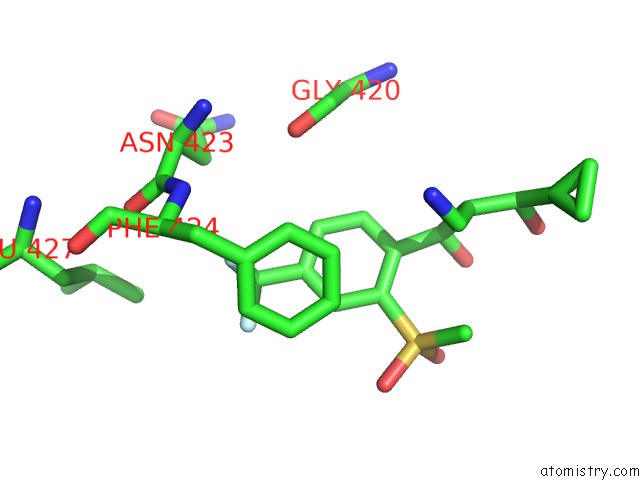
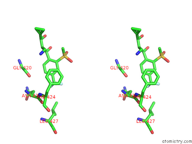
Fluorine binding site 1 out of 3 in 7x5u
Go back to
Fluorine binding site 1 out
of 3 in the Crystal Structure of Athppd-Diketonitrile Complex
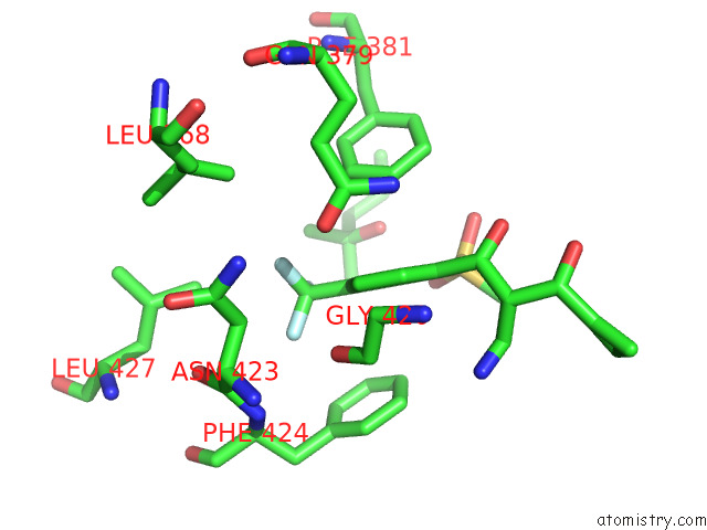
Mono view
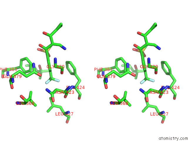
Stereo pair view
Mono view
Stereo pair view
A full contact list of Fluorine with other atoms in the F binding
site number 1 of Crystal Structure of Athppd-Diketonitrile Complex within 5.0Å range:
|
Fluorine binding site 2 out of 3 in 7x5u
Go back to
Fluorine binding site 2 out
of 3 in the Crystal Structure of Athppd-Diketonitrile Complex
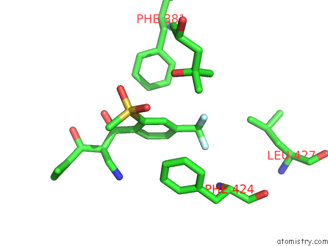
Mono view
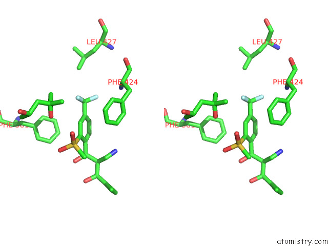
Stereo pair view
Mono view
Stereo pair view
A full contact list of Fluorine with other atoms in the F binding
site number 2 of Crystal Structure of Athppd-Diketonitrile Complex within 5.0Å range:
|
Fluorine binding site 3 out of 3 in 7x5u
Go back to
Fluorine binding site 3 out
of 3 in the Crystal Structure of Athppd-Diketonitrile Complex
Mono view
Stereo pair view
Mono view
Stereo pair view
A full contact list of Fluorine with other atoms in the F binding
site number 3 of Crystal Structure of Athppd-Diketonitrile Complex within 5.0Å range:
|
Reference:
J.Dong,
J.Dong,
X.H.Yu,
Y.C.Yan,
J.X.Nan,
B.He,
B.Q.Ye,
W.C.Yang,
H.Y.Lin,
G.F.Yang.
Structural Insights of 4-Hydrophenylpyruvate Dioxygenase Inhibition By Structurally Diverse Small Molecules Adv Agrochem 2022.
ISSN: ESSN 2773-2371
DOI: 10.1016/J.AAC.2022.10.002
Page generated: Fri Aug 2 15:20:44 2024
ISSN: ESSN 2773-2371
DOI: 10.1016/J.AAC.2022.10.002
Last articles
Zn in 9JYWZn in 9IR4
Zn in 9IR3
Zn in 9GMX
Zn in 9GMW
Zn in 9JEJ
Zn in 9ERF
Zn in 9ERE
Zn in 9EGV
Zn in 9EGW