Fluorine »
PDB 7y4g-7yzq »
7yxn »
Fluorine in PDB 7yxn: Crystal Structure of Wt ANCGR2-Lbd Bound to Dexamethasone and Shp Coregulator Fragment
Protein crystallography data
The structure of Crystal Structure of Wt ANCGR2-Lbd Bound to Dexamethasone and Shp Coregulator Fragment, PDB code: 7yxn
was solved by
A.Jimenez-Panizo,
E.Estebanez-Perpina,
P.Fuentes-Prior,
with X-Ray Crystallography technique. A brief refinement statistics is given in the table below:
| Resolution Low / High (Å) | 94.81 / 2.46 |
| Space group | P 61 |
| Cell size a, b, c (Å), α, β, γ (°) | 109.479, 109.479, 137.781, 90, 90, 120 |
| R / Rfree (%) | 21.6 / 24.7 |
Fluorine Binding Sites:
The binding sites of Fluorine atom in the Crystal Structure of Wt ANCGR2-Lbd Bound to Dexamethasone and Shp Coregulator Fragment
(pdb code 7yxn). This binding sites where shown within
5.0 Angstroms radius around Fluorine atom.
In total 2 binding sites of Fluorine where determined in the Crystal Structure of Wt ANCGR2-Lbd Bound to Dexamethasone and Shp Coregulator Fragment, PDB code: 7yxn:
Jump to Fluorine binding site number: 1; 2;
In total 2 binding sites of Fluorine where determined in the Crystal Structure of Wt ANCGR2-Lbd Bound to Dexamethasone and Shp Coregulator Fragment, PDB code: 7yxn:
Jump to Fluorine binding site number: 1; 2;
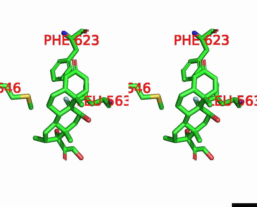
Fluorine binding site 1 out of 2 in 7yxn
Go back to
Fluorine binding site 1 out
of 2 in the Crystal Structure of Wt ANCGR2-Lbd Bound to Dexamethasone and Shp Coregulator Fragment

Mono view

Stereo pair view
Mono view
Stereo pair view
A full contact list of Fluorine with other atoms in the F binding
site number 1 of Crystal Structure of Wt ANCGR2-Lbd Bound to Dexamethasone and Shp Coregulator Fragment within 5.0Å range:
|
Fluorine binding site 2 out of 2 in 7yxn
Go back to
Fluorine binding site 2 out
of 2 in the Crystal Structure of Wt ANCGR2-Lbd Bound to Dexamethasone and Shp Coregulator Fragment

Mono view
Stereo pair view
Mono view
Stereo pair view
A full contact list of Fluorine with other atoms in the F binding
site number 2 of Crystal Structure of Wt ANCGR2-Lbd Bound to Dexamethasone and Shp Coregulator Fragment within 5.0Å range:
|
Reference:
A.Jimenez-Panizo,
A.Alegre-Marti,
T.T.Tettey,
G.Fettweis,
M.Abella,
R.Anton,
T.A.Johnson,
S.Kim,
R.L.Schiltz,
I.Nunez-Barrios,
J.Font-Diaz,
C.Caelles,
A.F.Valledor,
P.Perez,
A.M.Rojas,
J.Fernandez-Recio,
D.M.Presman,
G.L.Hager,
P.Fuentes-Prior,
E.Estebanez-Perpina.
The Multivalency of the Glucocorticoid Receptor Ligand-Binding Domain Explains Its Manifold Physiological Activities. Nucleic Acids Res. 2022.
ISSN: ESSN 1362-4962
PubMed: 36464162
DOI: 10.1093/NAR/GKAC1119
Page generated: Wed Jul 16 02:18:54 2025
ISSN: ESSN 1362-4962
PubMed: 36464162
DOI: 10.1093/NAR/GKAC1119
Last articles
Fe in 2YXOFe in 2YRS
Fe in 2YXC
Fe in 2YNM
Fe in 2YVJ
Fe in 2YP1
Fe in 2YU2
Fe in 2YU1
Fe in 2YQB
Fe in 2YOO