Fluorine »
PDB 8gky-8hej »
8h57 »
Fluorine in PDB 8h57: Crystal Structure of Sars-Cov-2 Main Protease (Mpro) A193P Mutant in Complex with Inhibitor Nirmatrelvir
Enzymatic activity of Crystal Structure of Sars-Cov-2 Main Protease (Mpro) A193P Mutant in Complex with Inhibitor Nirmatrelvir
All present enzymatic activity of Crystal Structure of Sars-Cov-2 Main Protease (Mpro) A193P Mutant in Complex with Inhibitor Nirmatrelvir:
3.4.22.69;
3.4.22.69;
Protein crystallography data
The structure of Crystal Structure of Sars-Cov-2 Main Protease (Mpro) A193P Mutant in Complex with Inhibitor Nirmatrelvir, PDB code: 8h57
was solved by
M.Lin,
X.Liu,
with X-Ray Crystallography technique. A brief refinement statistics is given in the table below:
| Resolution Low / High (Å) | 27.80 / 1.55 |
| Space group | P 21 21 2 |
| Cell size a, b, c (Å), α, β, γ (°) | 45.399, 63.974, 105.469, 90, 90, 90 |
| R / Rfree (%) | 17.7 / 20.4 |
Fluorine Binding Sites:
The binding sites of Fluorine atom in the Crystal Structure of Sars-Cov-2 Main Protease (Mpro) A193P Mutant in Complex with Inhibitor Nirmatrelvir
(pdb code 8h57). This binding sites where shown within
5.0 Angstroms radius around Fluorine atom.
In total 3 binding sites of Fluorine where determined in the Crystal Structure of Sars-Cov-2 Main Protease (Mpro) A193P Mutant in Complex with Inhibitor Nirmatrelvir, PDB code: 8h57:
Jump to Fluorine binding site number: 1; 2; 3;
In total 3 binding sites of Fluorine where determined in the Crystal Structure of Sars-Cov-2 Main Protease (Mpro) A193P Mutant in Complex with Inhibitor Nirmatrelvir, PDB code: 8h57:
Jump to Fluorine binding site number: 1; 2; 3;
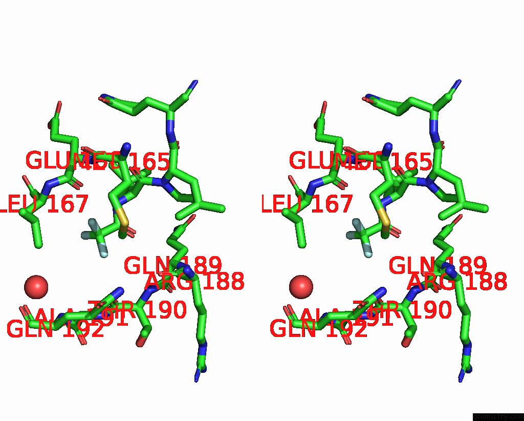
Fluorine binding site 1 out of 3 in 8h57
Go back to
Fluorine binding site 1 out
of 3 in the Crystal Structure of Sars-Cov-2 Main Protease (Mpro) A193P Mutant in Complex with Inhibitor Nirmatrelvir
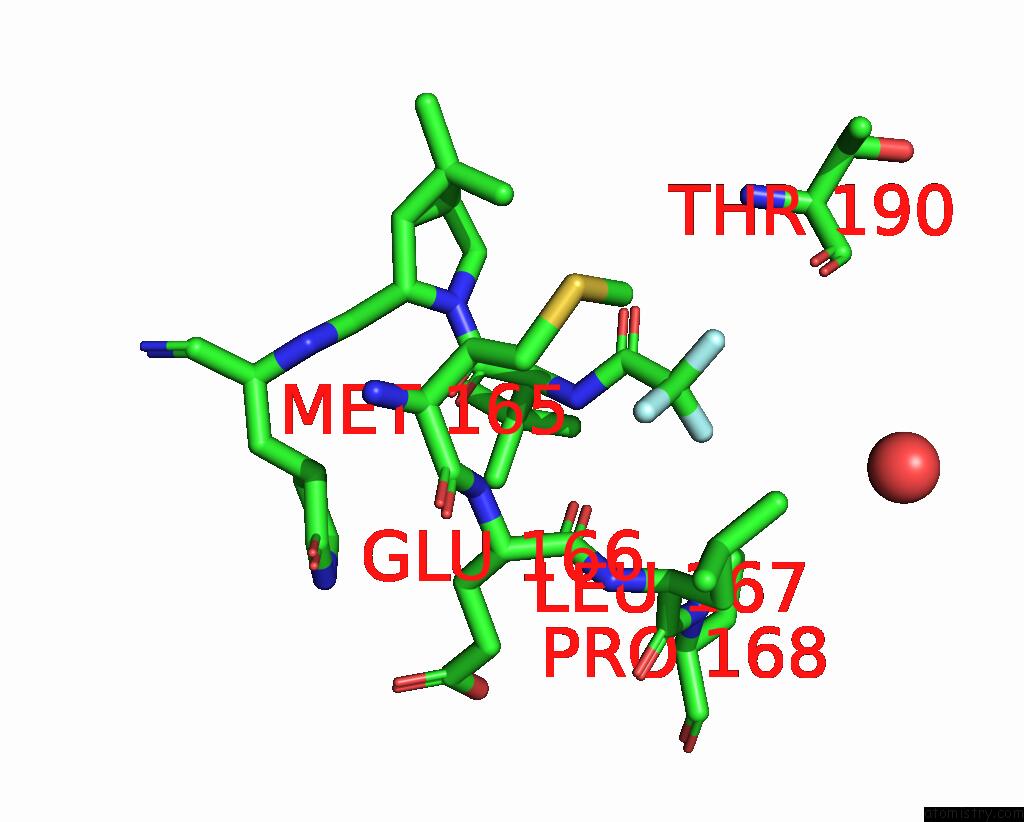
Mono view
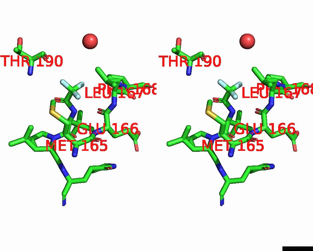
Stereo pair view
Mono view
Stereo pair view
A full contact list of Fluorine with other atoms in the F binding
site number 1 of Crystal Structure of Sars-Cov-2 Main Protease (Mpro) A193P Mutant in Complex with Inhibitor Nirmatrelvir within 5.0Å range:
|
Fluorine binding site 2 out of 3 in 8h57
Go back to
Fluorine binding site 2 out
of 3 in the Crystal Structure of Sars-Cov-2 Main Protease (Mpro) A193P Mutant in Complex with Inhibitor Nirmatrelvir
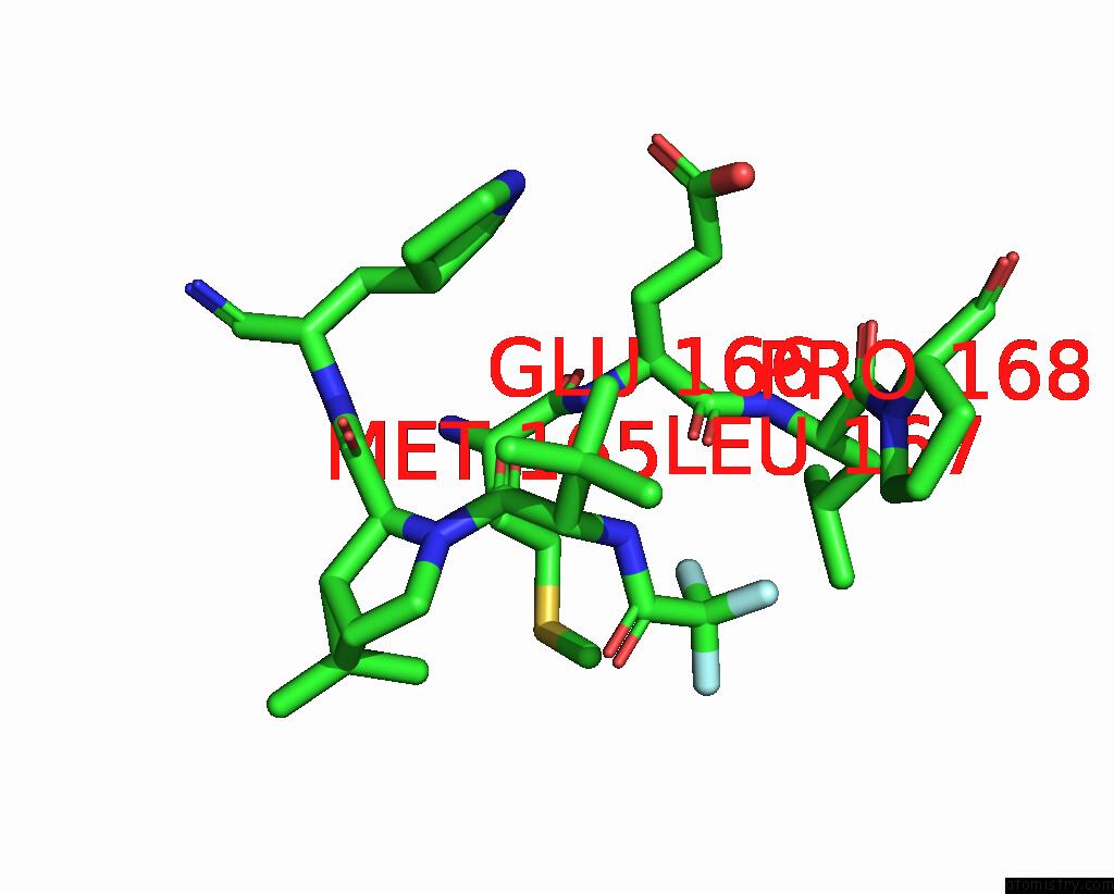
Mono view
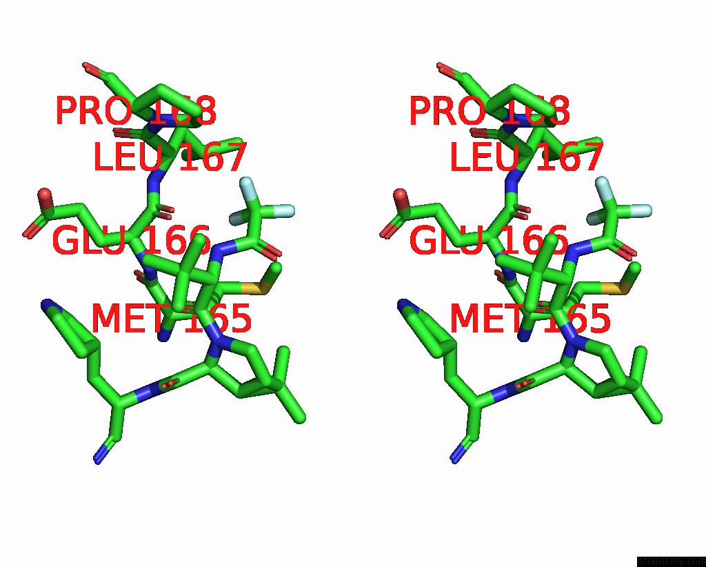
Stereo pair view
Mono view
Stereo pair view
A full contact list of Fluorine with other atoms in the F binding
site number 2 of Crystal Structure of Sars-Cov-2 Main Protease (Mpro) A193P Mutant in Complex with Inhibitor Nirmatrelvir within 5.0Å range:
|
Fluorine binding site 3 out of 3 in 8h57
Go back to
Fluorine binding site 3 out
of 3 in the Crystal Structure of Sars-Cov-2 Main Protease (Mpro) A193P Mutant in Complex with Inhibitor Nirmatrelvir

Mono view
Stereo pair view
Mono view
Stereo pair view
A full contact list of Fluorine with other atoms in the F binding
site number 3 of Crystal Structure of Sars-Cov-2 Main Protease (Mpro) A193P Mutant in Complex with Inhibitor Nirmatrelvir within 5.0Å range:
|
Reference:
Y.Duan,
H.Zhou,
X.Liu,
S.Iketani,
M.Lin,
X.Zhang,
Q.Bian,
H.Wang,
H.Sun,
S.J.Hong,
B.Culbertson,
H.Mohri,
M.I.Luck,
Y.Zhu,
X.Liu,
Y.Lu,
X.Yang,
K.Yang,
Y.Sabo,
A.Chavez,
S.P.Goff,
Z.Rao,
D.D.Ho,
H.Yang.
Molecular Mechanisms of Sars-Cov-2 Resistance to Nirmatrelvir. Nature 2023.
ISSN: ESSN 1476-4687
PubMed: 37696289
DOI: 10.1038/S41586-023-06609-0
Page generated: Wed Jul 16 05:10:44 2025
ISSN: ESSN 1476-4687
PubMed: 37696289
DOI: 10.1038/S41586-023-06609-0
Last articles
Fe in 1CJXFe in 1CL6
Fe in 1CK6
Fe in 1CIO
Fe in 1CIK
Fe in 1CIH
Fe in 1CIG
Fe in 1CIF
Fe in 1CI6
Fe in 1CH4