Fluorine in PDB 8hbk: The Crystal Structure of Sars-Cov-2 3CL Protease in Complex with Ensitrelvir
Enzymatic activity of The Crystal Structure of Sars-Cov-2 3CL Protease in Complex with Ensitrelvir
All present enzymatic activity of The Crystal Structure of Sars-Cov-2 3CL Protease in Complex with Ensitrelvir:
3.4.22.69;
3.4.22.69;
Protein crystallography data
The structure of The Crystal Structure of Sars-Cov-2 3CL Protease in Complex with Ensitrelvir, PDB code: 8hbk
was solved by
M.Lin,
with X-Ray Crystallography technique. A brief refinement statistics is given in the table below:
| Resolution Low / High (Å) | 41.26 / 1.80 |
| Space group | C 1 2 1 |
| Cell size a, b, c (Å), α, β, γ (°) | 97.093, 82.528, 51.526, 90, 115.13, 90 |
| R / Rfree (%) | 19.1 / 23.2 |
Other elements in 8hbk:
The structure of The Crystal Structure of Sars-Cov-2 3CL Protease in Complex with Ensitrelvir also contains other interesting chemical elements:
| Chlorine | (Cl) | 1 atom |
Fluorine Binding Sites:
The binding sites of Fluorine atom in the The Crystal Structure of Sars-Cov-2 3CL Protease in Complex with Ensitrelvir
(pdb code 8hbk). This binding sites where shown within
5.0 Angstroms radius around Fluorine atom.
In total 3 binding sites of Fluorine where determined in the The Crystal Structure of Sars-Cov-2 3CL Protease in Complex with Ensitrelvir, PDB code: 8hbk:
Jump to Fluorine binding site number: 1; 2; 3;
In total 3 binding sites of Fluorine where determined in the The Crystal Structure of Sars-Cov-2 3CL Protease in Complex with Ensitrelvir, PDB code: 8hbk:
Jump to Fluorine binding site number: 1; 2; 3;
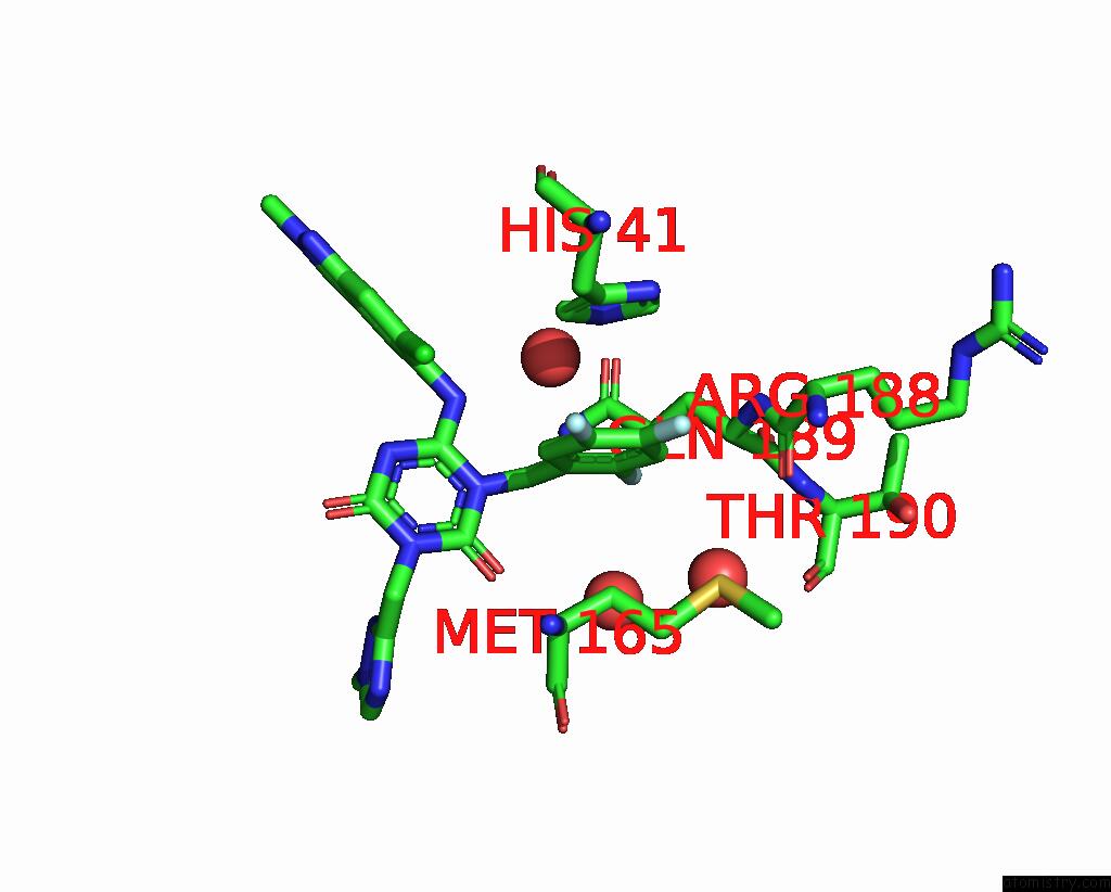
Fluorine binding site 1 out of 3 in 8hbk
Go back to
Fluorine binding site 1 out
of 3 in the The Crystal Structure of Sars-Cov-2 3CL Protease in Complex with Ensitrelvir
Mono view
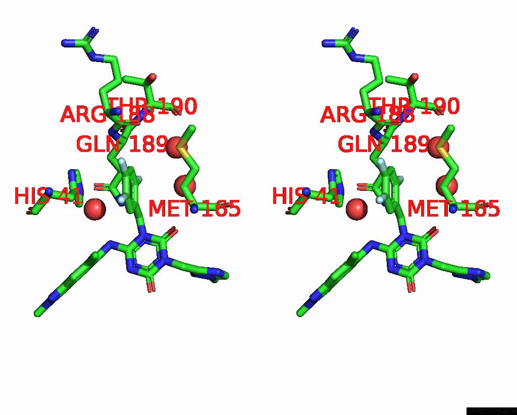
Stereo pair view
Mono view
Stereo pair view
A full contact list of Fluorine with other atoms in the F binding
site number 1 of The Crystal Structure of Sars-Cov-2 3CL Protease in Complex with Ensitrelvir within 5.0Å range:
|
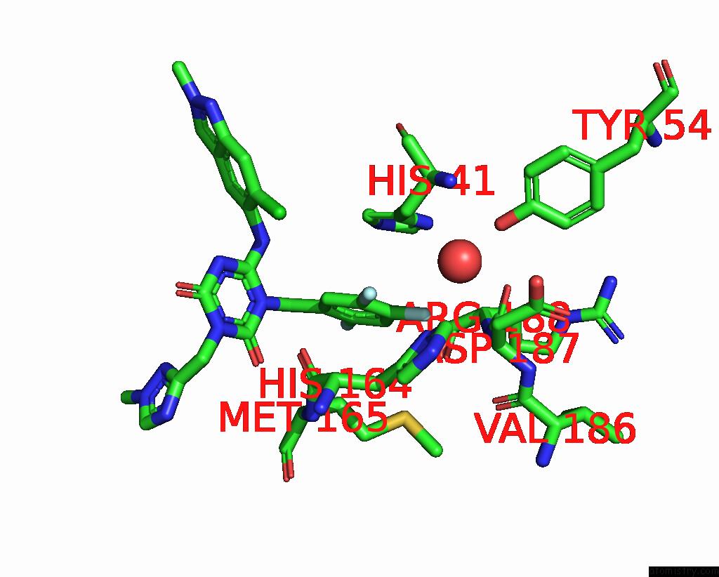
Fluorine binding site 2 out of 3 in 8hbk
Go back to
Fluorine binding site 2 out
of 3 in the The Crystal Structure of Sars-Cov-2 3CL Protease in Complex with Ensitrelvir
Mono view

Stereo pair view
Mono view
Stereo pair view
A full contact list of Fluorine with other atoms in the F binding
site number 2 of The Crystal Structure of Sars-Cov-2 3CL Protease in Complex with Ensitrelvir within 5.0Å range:
|
Fluorine binding site 3 out of 3 in 8hbk
Go back to
Fluorine binding site 3 out
of 3 in the The Crystal Structure of Sars-Cov-2 3CL Protease in Complex with Ensitrelvir

Mono view

Stereo pair view
Mono view
Stereo pair view
A full contact list of Fluorine with other atoms in the F binding
site number 3 of The Crystal Structure of Sars-Cov-2 3CL Protease in Complex with Ensitrelvir within 5.0Å range:
|
Reference:
Y.Duan,
H.Zhou,
X.Liu,
S.Iketani,
M.Lin,
X.Zhang,
Q.Bian,
H.Wang,
H.Sun,
S.J.Hong,
B.Culbertson,
H.Mohri,
M.I.Luck,
Y.Zhu,
X.Liu,
Y.Lu,
X.Yang,
K.Yang,
Y.Sabo,
A.Chavez,
S.P.Goff,
Z.Rao,
D.D.Ho,
H.Yang.
Molecular Mechanisms of Sars-Cov-2 Resistance to Nirmatrelvir. Nature 2023.
ISSN: ESSN 1476-4687
PubMed: 37696289
DOI: 10.1038/S41586-023-06609-0
Page generated: Fri Aug 2 20:06:54 2024
ISSN: ESSN 1476-4687
PubMed: 37696289
DOI: 10.1038/S41586-023-06609-0
Last articles
Zn in 9MJ5Zn in 9HNW
Zn in 9G0L
Zn in 9FNE
Zn in 9DZN
Zn in 9E0I
Zn in 9D32
Zn in 9DAK
Zn in 8ZXC
Zn in 8ZUF