Fluorine »
PDB 8pgx-8q0v »
8pl0 »
Fluorine in PDB 8pl0: Thioredoxin Glutathione Reductase of Schistosoma Mansoni Fragment Screen Hit 1.
Enzymatic activity of Thioredoxin Glutathione Reductase of Schistosoma Mansoni Fragment Screen Hit 1.
All present enzymatic activity of Thioredoxin Glutathione Reductase of Schistosoma Mansoni Fragment Screen Hit 1.:
1.6.4.5;
1.6.4.5;
Protein crystallography data
The structure of Thioredoxin Glutathione Reductase of Schistosoma Mansoni Fragment Screen Hit 1., PDB code: 8pl0
was solved by
L.Ribeiro,
B.O.Montoya,
J.T.Moreira-Filho,
S.Bowyer,
A.Verma,
B.J.Neves,
R.J.Owens,
C.H.Andrade,
F.P.Silva-Jr,
N.Furnham,
with X-Ray Crystallography technique. A brief refinement statistics is given in the table below:
| Resolution Low / High (Å) | 29.67 / 1.70 |
| Space group | P 1 21 1 |
| Cell size a, b, c (Å), α, β, γ (°) | 62.626, 103.919, 134.14, 90, 91.97, 90 |
| R / Rfree (%) | 18.9 / 21.2 |
Fluorine Binding Sites:
The binding sites of Fluorine atom in the Thioredoxin Glutathione Reductase of Schistosoma Mansoni Fragment Screen Hit 1.
(pdb code 8pl0). This binding sites where shown within
5.0 Angstroms radius around Fluorine atom.
In total only one binding site of Fluorine was determined in the Thioredoxin Glutathione Reductase of Schistosoma Mansoni Fragment Screen Hit 1., PDB code: 8pl0:
In total only one binding site of Fluorine was determined in the Thioredoxin Glutathione Reductase of Schistosoma Mansoni Fragment Screen Hit 1., PDB code: 8pl0:
Fluorine binding site 1 out of 1 in 8pl0
Go back to
Fluorine binding site 1 out
of 1 in the Thioredoxin Glutathione Reductase of Schistosoma Mansoni Fragment Screen Hit 1.
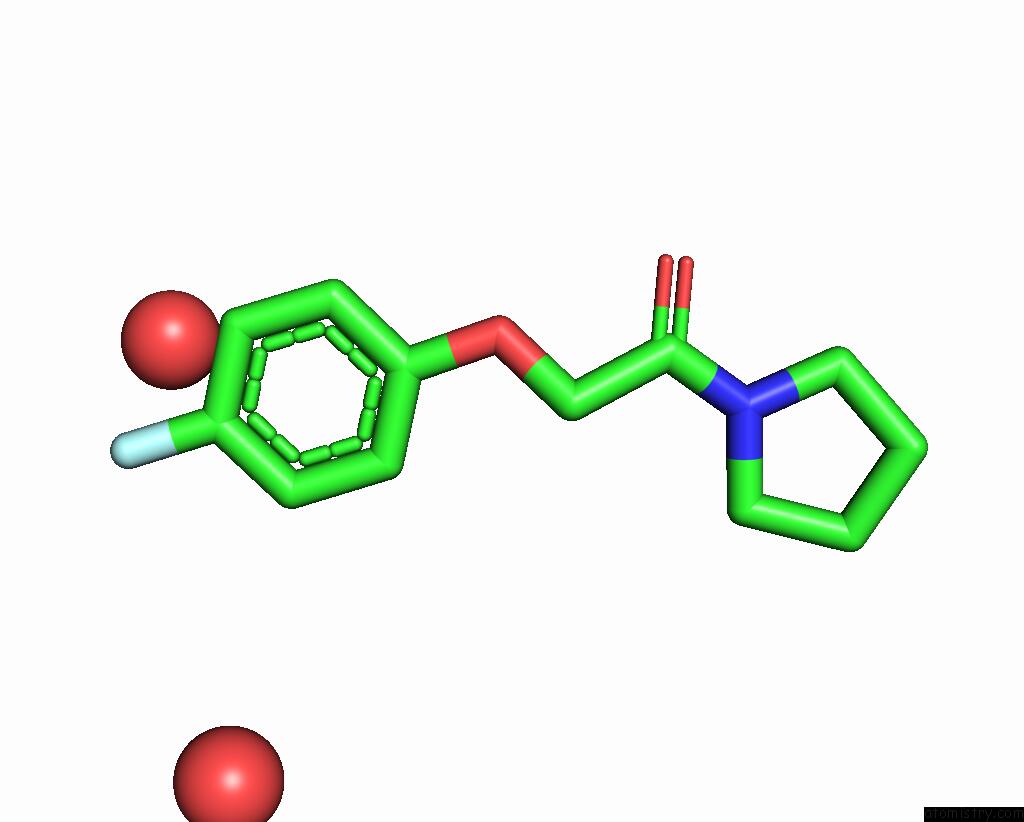
Mono view
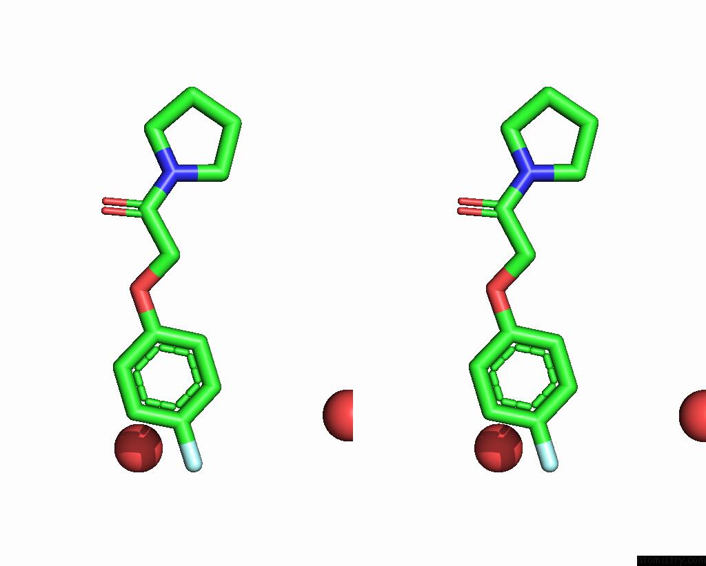
Stereo pair view
Mono view
Stereo pair view
A full contact list of Fluorine with other atoms in the F binding
site number 1 of Thioredoxin Glutathione Reductase of Schistosoma Mansoni Fragment Screen Hit 1. within 5.0Å range:
|
Reference:
L.R.De Souza Neto,
B.O.Montoya,
J.Brandao-Neto,
A.Verma,
S.Bowyer,
J.T.Moreira-Filho,
R.F.Dantas,
B.J.Neves,
C.H.Andrade,
F.Von Delft,
R.J.Owens,
N.Furnham,
F.P.Silva-Jr.
Fragment Library Screening By X-Ray Crystallography and Binding Site Analysis on Thioredoxin Glutathione Reductase of Schistosoma Mansoni. Sci Rep V. 14 1582 2024.
ISSN: ESSN 2045-2322
PubMed: 38238498
DOI: 10.1038/S41598-024-52018-2
Page generated: Wed Jul 16 06:40:23 2025
ISSN: ESSN 2045-2322
PubMed: 38238498
DOI: 10.1038/S41598-024-52018-2
Last articles
Fe in 2YXOFe in 2YRS
Fe in 2YXC
Fe in 2YNM
Fe in 2YVJ
Fe in 2YP1
Fe in 2YU2
Fe in 2YU1
Fe in 2YQB
Fe in 2YOO