Fluorine »
PDB 8pgx-8q0v »
8pp0 »
Fluorine in PDB 8pp0: Crystal Structure of Retinoic Acid Receptor Alpha (Rxra) in Complexed with JP147
Protein crystallography data
The structure of Crystal Structure of Retinoic Acid Receptor Alpha (Rxra) in Complexed with JP147, PDB code: 8pp0
was solved by
A.Chaikuad,
J.Pollinger,
D.Merk,
S.Knapp,
Structural Genomics Consortium(Sgc),
with X-Ray Crystallography technique. A brief refinement statistics is given in the table below:
| Resolution Low / High (Å) | 42.85 / 1.90 |
| Space group | P 43 21 2 |
| Cell size a, b, c (Å), α, β, γ (°) | 65.648, 65.648, 110.816, 90, 90, 90 |
| R / Rfree (%) | 18 / 22.3 |
Fluorine Binding Sites:
The binding sites of Fluorine atom in the Crystal Structure of Retinoic Acid Receptor Alpha (Rxra) in Complexed with JP147
(pdb code 8pp0). This binding sites where shown within
5.0 Angstroms radius around Fluorine atom.
In total 3 binding sites of Fluorine where determined in the Crystal Structure of Retinoic Acid Receptor Alpha (Rxra) in Complexed with JP147, PDB code: 8pp0:
Jump to Fluorine binding site number: 1; 2; 3;
In total 3 binding sites of Fluorine where determined in the Crystal Structure of Retinoic Acid Receptor Alpha (Rxra) in Complexed with JP147, PDB code: 8pp0:
Jump to Fluorine binding site number: 1; 2; 3;
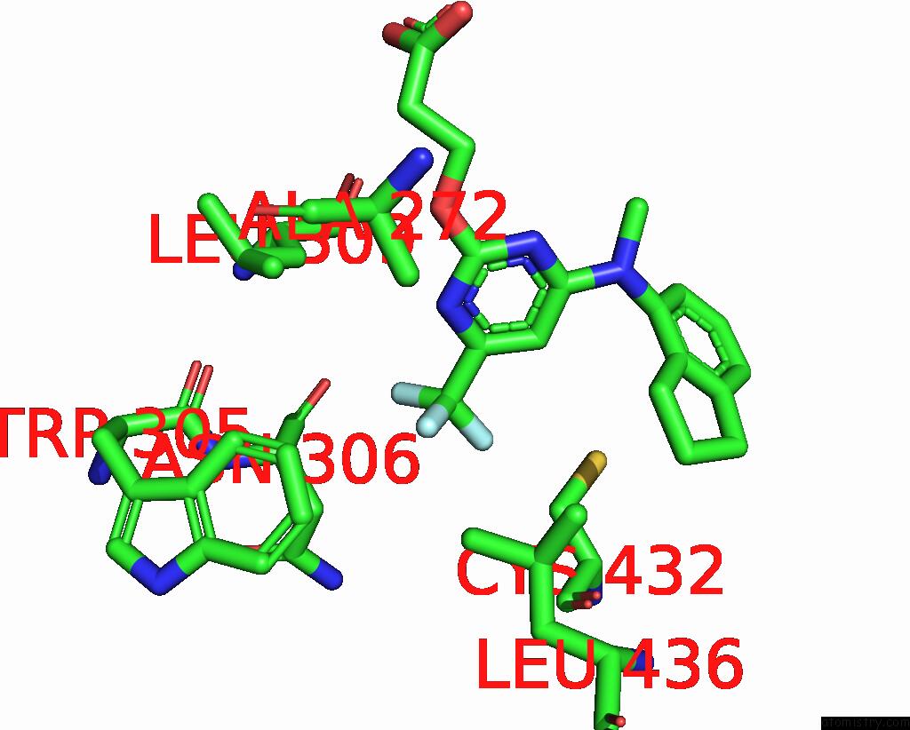
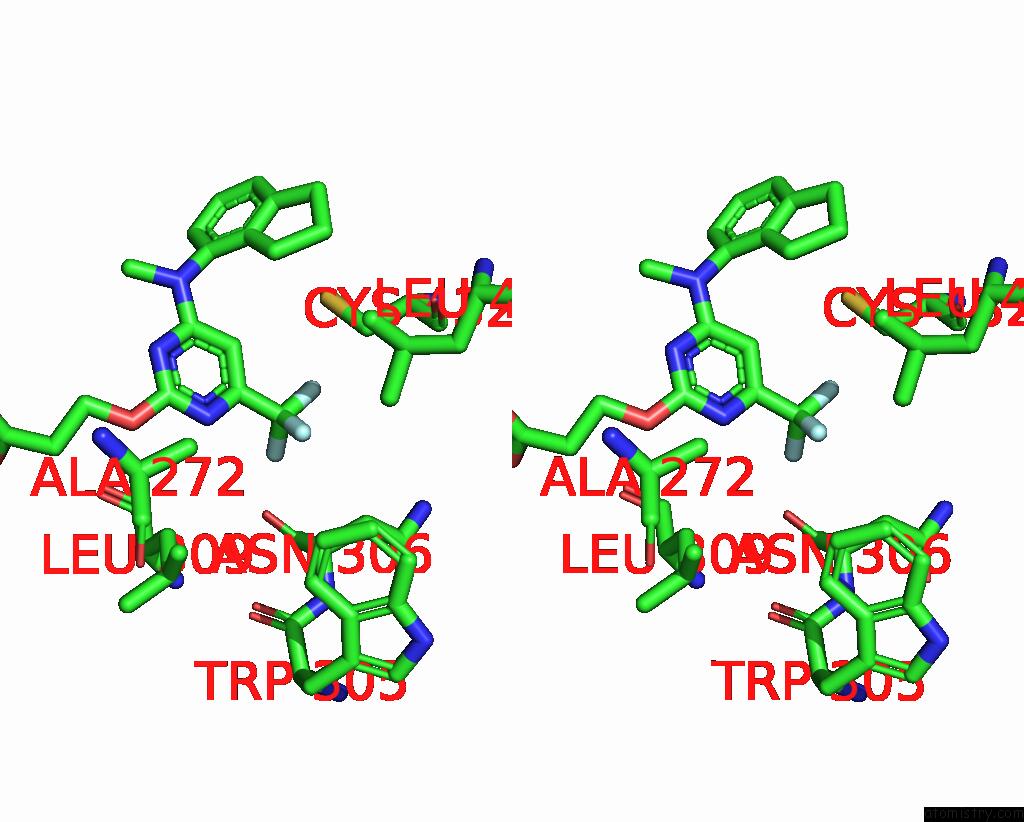
Fluorine binding site 1 out of 3 in 8pp0
Go back to
Fluorine binding site 1 out
of 3 in the Crystal Structure of Retinoic Acid Receptor Alpha (Rxra) in Complexed with JP147
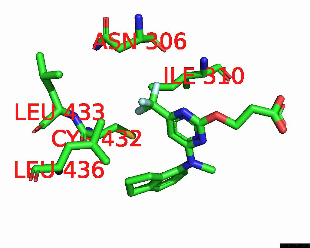
Mono view
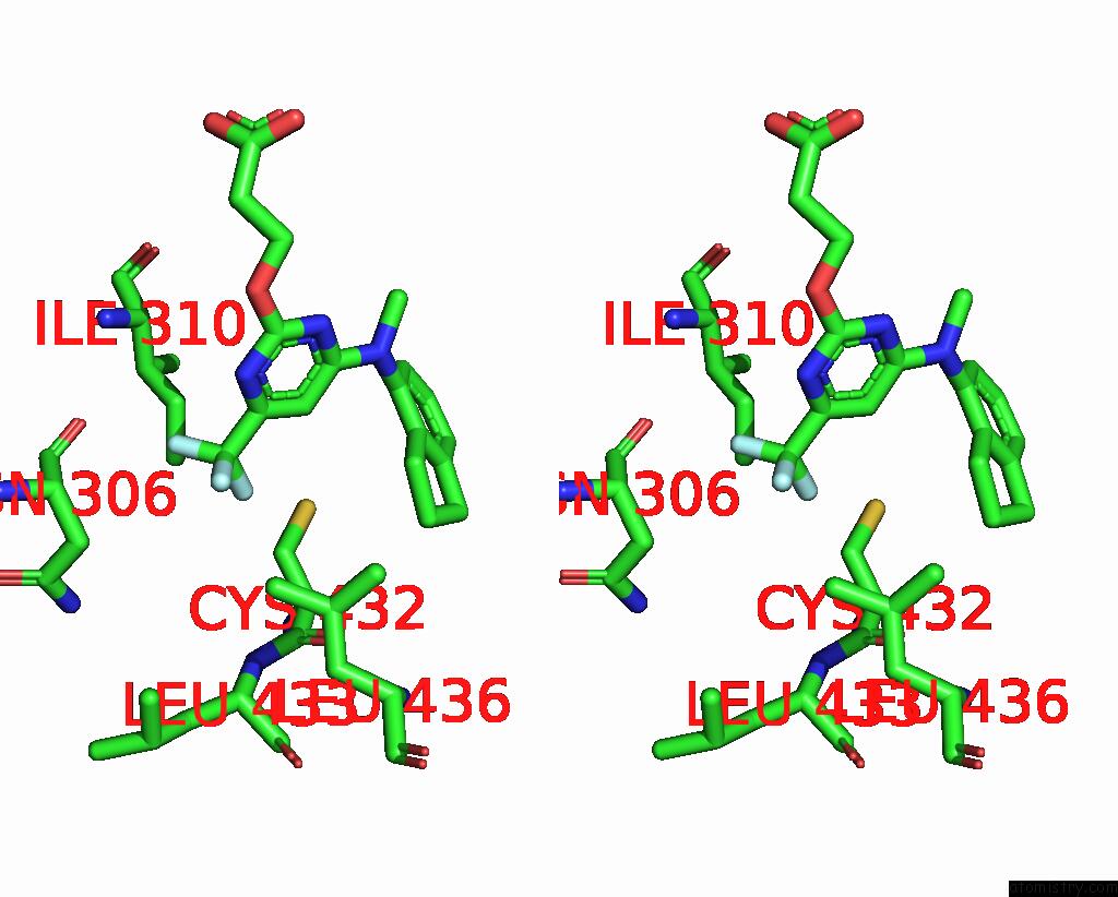
Stereo pair view
Mono view
Stereo pair view
A full contact list of Fluorine with other atoms in the F binding
site number 1 of Crystal Structure of Retinoic Acid Receptor Alpha (Rxra) in Complexed with JP147 within 5.0Å range:
|
Fluorine binding site 2 out of 3 in 8pp0
Go back to
Fluorine binding site 2 out
of 3 in the Crystal Structure of Retinoic Acid Receptor Alpha (Rxra) in Complexed with JP147
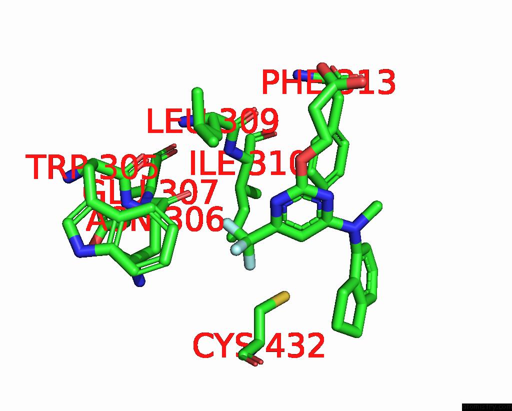
Mono view
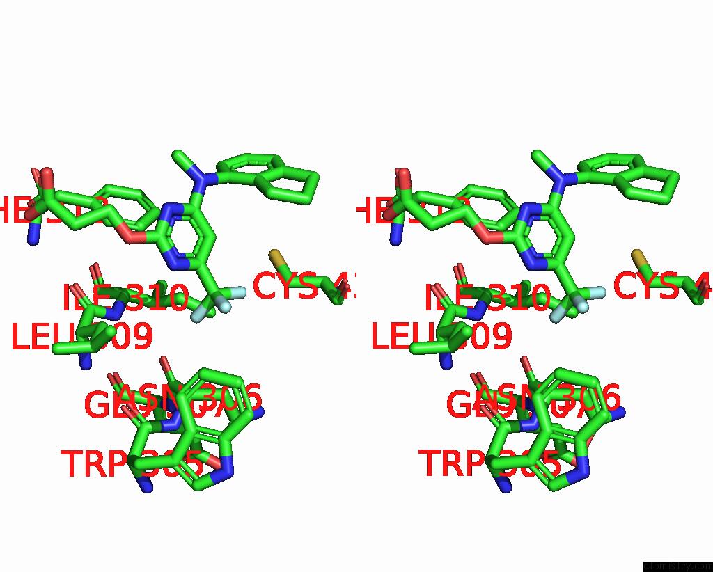
Stereo pair view
Mono view
Stereo pair view
A full contact list of Fluorine with other atoms in the F binding
site number 2 of Crystal Structure of Retinoic Acid Receptor Alpha (Rxra) in Complexed with JP147 within 5.0Å range:
|
Fluorine binding site 3 out of 3 in 8pp0
Go back to
Fluorine binding site 3 out
of 3 in the Crystal Structure of Retinoic Acid Receptor Alpha (Rxra) in Complexed with JP147
Mono view
Stereo pair view
Mono view
Stereo pair view
A full contact list of Fluorine with other atoms in the F binding
site number 3 of Crystal Structure of Retinoic Acid Receptor Alpha (Rxra) in Complexed with JP147 within 5.0Å range:
|
Reference:
M.Lewandowski,
M.Carmina,
L.Knumann,
M.Sai,
S.Willems,
T.Kasch,
J.Pollinger,
S.Knapp,
J.A.Marschner,
A.Chaikuad,
D.Merk.
Structure-Guided Design of A Highly Potent Partial Rxr Agonist with Superior Physicochemical Properties. J.Med.Chem. 2024.
ISSN: ISSN 0022-2623
PubMed: 38237049
DOI: 10.1021/ACS.JMEDCHEM.3C02095
Page generated: Wed Jul 16 06:42:42 2025
ISSN: ISSN 0022-2623
PubMed: 38237049
DOI: 10.1021/ACS.JMEDCHEM.3C02095
Last articles
Fe in 2YXOFe in 2YRS
Fe in 2YXC
Fe in 2YNM
Fe in 2YVJ
Fe in 2YP1
Fe in 2YU2
Fe in 2YU1
Fe in 2YQB
Fe in 2YOO