Fluorine »
PDB 8pgx-8q0v »
8pqi »
Fluorine in PDB 8pqi: Pdgfra T674I Mutant Kinase Domain in Complex with Avapritinib Derivative 9
Enzymatic activity of Pdgfra T674I Mutant Kinase Domain in Complex with Avapritinib Derivative 9
All present enzymatic activity of Pdgfra T674I Mutant Kinase Domain in Complex with Avapritinib Derivative 9:
2.7.10.1;
2.7.10.1;
Protein crystallography data
The structure of Pdgfra T674I Mutant Kinase Domain in Complex with Avapritinib Derivative 9, PDB code: 8pqi
was solved by
A.Teuber,
S.Kleinboelting,
M.P.Mueller,
D.Rauh,
with X-Ray Crystallography technique. A brief refinement statistics is given in the table below:
| Resolution Low / High (Å) | 46.63 / 2.60 |
| Space group | P 21 21 21 |
| Cell size a, b, c (Å), α, β, γ (°) | 52.39, 74.04, 102.26, 90, 90, 90 |
| R / Rfree (%) | 23.1 / 25.4 |
Fluorine Binding Sites:
The binding sites of Fluorine atom in the Pdgfra T674I Mutant Kinase Domain in Complex with Avapritinib Derivative 9
(pdb code 8pqi). This binding sites where shown within
5.0 Angstroms radius around Fluorine atom.
In total only one binding site of Fluorine was determined in the Pdgfra T674I Mutant Kinase Domain in Complex with Avapritinib Derivative 9, PDB code: 8pqi:
In total only one binding site of Fluorine was determined in the Pdgfra T674I Mutant Kinase Domain in Complex with Avapritinib Derivative 9, PDB code: 8pqi:
Fluorine binding site 1 out of 1 in 8pqi
Go back to
Fluorine binding site 1 out
of 1 in the Pdgfra T674I Mutant Kinase Domain in Complex with Avapritinib Derivative 9
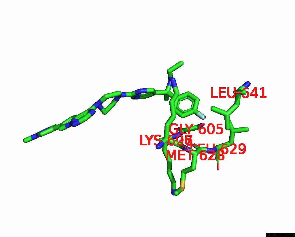
Mono view
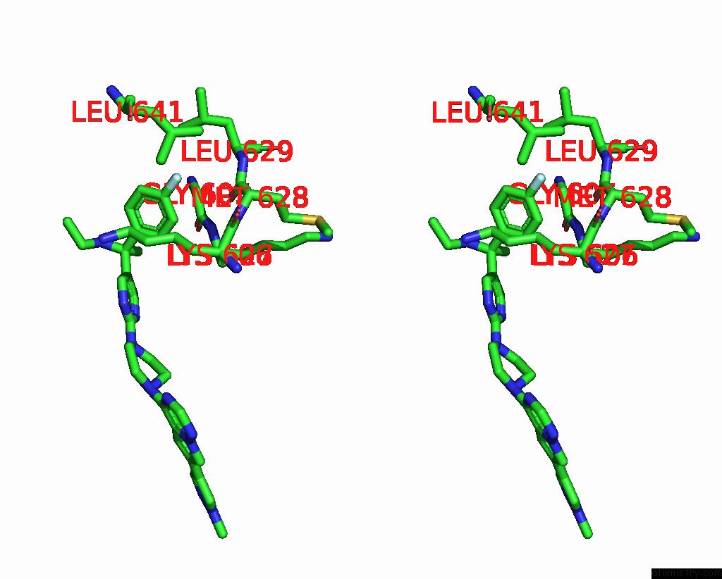
Stereo pair view
Mono view
Stereo pair view
A full contact list of Fluorine with other atoms in the F binding
site number 1 of Pdgfra T674I Mutant Kinase Domain in Complex with Avapritinib Derivative 9 within 5.0Å range:
|
Reference:
A.Teuber,
T.Schulz,
B.S.Fletcher,
R.Gontla,
T.Muehlenberg,
M.-L.Zischinsky,
J.Niggenaber,
J.Weisner,
S.B.Kleinboelting,
J.Lategahn,
S.Sievers,
M.P.Mueller,
S.Bauer,
D.Rauh.
Avapritinib-Based Sar Studies Reveal A Binding Pocket in Kit and Pdgfra Nat Commun 2023.
ISSN: ESSN 2041-1723
DOI: 10.1038/S41467-023-44376-8
Page generated: Wed Jul 16 06:43:54 2025
ISSN: ESSN 2041-1723
DOI: 10.1038/S41467-023-44376-8
Last articles
Fe in 2YXOFe in 2YRS
Fe in 2YXC
Fe in 2YNM
Fe in 2YVJ
Fe in 2YP1
Fe in 2YU2
Fe in 2YU1
Fe in 2YQB
Fe in 2YOO