Fluorine »
PDB 8smc-8t4v »
8t2h »
Fluorine in PDB 8t2h: DYRK1A Complex with DYR530
Enzymatic activity of DYRK1A Complex with DYR530
Protein crystallography data
The structure of DYRK1A Complex with DYR530, PDB code: 8t2h
was solved by
W.R.Montfort,
L.E.Basantes,
with X-Ray Crystallography technique. A brief refinement statistics is given in the table below:
| Resolution Low / High (Å) | 24.88 / 1.85 |
| Space group | P 21 21 21 |
| Cell size a, b, c (Å), α, β, γ (°) | 63.472, 83.213, 146.675, 90, 90, 90 |
| R / Rfree (%) | 16.3 / 21.3 |
Fluorine Binding Sites:
The binding sites of Fluorine atom in the DYRK1A Complex with DYR530
(pdb code 8t2h). This binding sites where shown within
5.0 Angstroms radius around Fluorine atom.
In total 2 binding sites of Fluorine where determined in the DYRK1A Complex with DYR530, PDB code: 8t2h:
Jump to Fluorine binding site number: 1; 2;
In total 2 binding sites of Fluorine where determined in the DYRK1A Complex with DYR530, PDB code: 8t2h:
Jump to Fluorine binding site number: 1; 2;
Fluorine binding site 1 out of 2 in 8t2h
Go back to
Fluorine binding site 1 out
of 2 in the DYRK1A Complex with DYR530
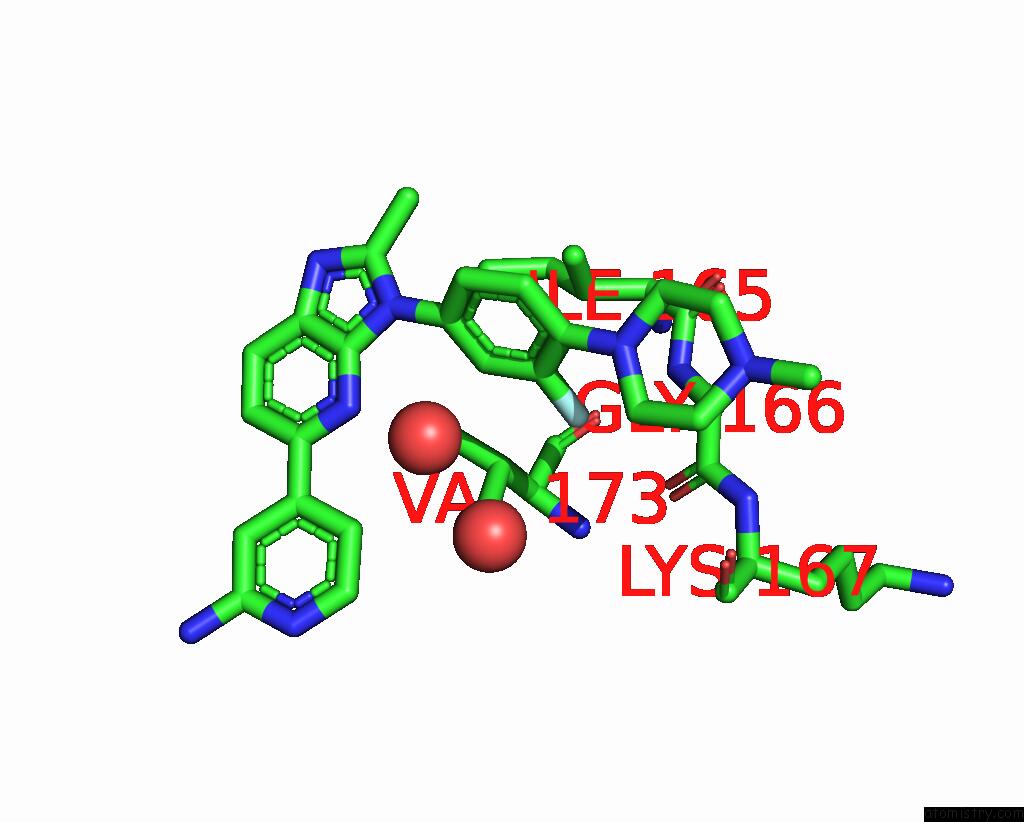
Mono view
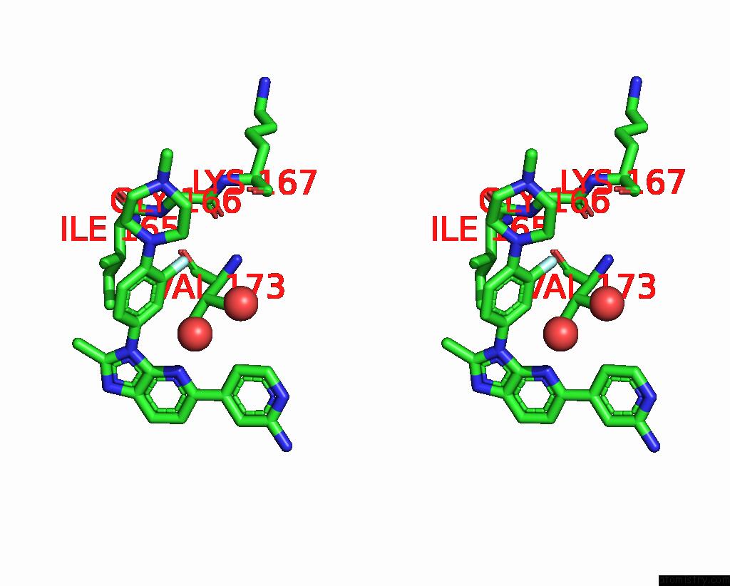
Stereo pair view
Mono view
Stereo pair view
A full contact list of Fluorine with other atoms in the F binding
site number 1 of DYRK1A Complex with DYR530 within 5.0Å range:
|
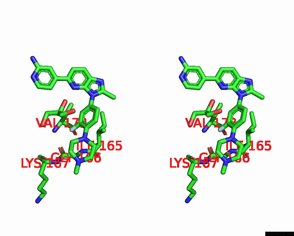
Fluorine binding site 2 out of 2 in 8t2h
Go back to
Fluorine binding site 2 out
of 2 in the DYRK1A Complex with DYR530
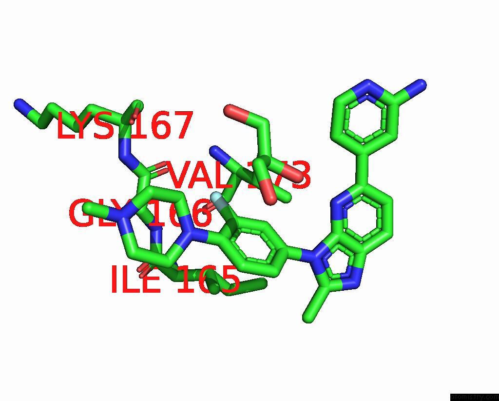
Mono view
Stereo pair view
Mono view
Stereo pair view
A full contact list of Fluorine with other atoms in the F binding
site number 2 of DYRK1A Complex with DYR530 within 5.0Å range:
|
Reference:
W.R.Montfort,
L.E.Basantes.
Discovery of DYR684, A Potent, Selective, Metabolically Stable, DYRK1A/B Protac Utilizing A Novel Cereblon Molecular Glue To Be Published.
Page generated: Wed Jul 16 08:29:17 2025
Last articles
Fe in 2YXOFe in 2YRS
Fe in 2YXC
Fe in 2YNM
Fe in 2YVJ
Fe in 2YP1
Fe in 2YU2
Fe in 2YU1
Fe in 2YQB
Fe in 2YOO