Fluorine »
PDB 9ars-9cdk »
9c7l »
Fluorine in PDB 9c7l: Crystal Structure of Pentalenene Synthase Variant F76A Complexed with 2-Fluorofarnesyl Diphosphate
Enzymatic activity of Crystal Structure of Pentalenene Synthase Variant F76A Complexed with 2-Fluorofarnesyl Diphosphate
All present enzymatic activity of Crystal Structure of Pentalenene Synthase Variant F76A Complexed with 2-Fluorofarnesyl Diphosphate:
4.2.3.7;
4.2.3.7;
Protein crystallography data
The structure of Crystal Structure of Pentalenene Synthase Variant F76A Complexed with 2-Fluorofarnesyl Diphosphate, PDB code: 9c7l
was solved by
R.Prem Kumar,
W.H.Ellenburg,
D.D.Oprian,
with X-Ray Crystallography technique. A brief refinement statistics is given in the table below:
| Resolution Low / High (Å) | 19.84 / 2.20 |
| Space group | P 63 |
| Cell size a, b, c (Å), α, β, γ (°) | 181.521, 181.521, 55.763, 90, 90, 120 |
| R / Rfree (%) | 18.5 / 21.3 |
Other elements in 9c7l:
The structure of Crystal Structure of Pentalenene Synthase Variant F76A Complexed with 2-Fluorofarnesyl Diphosphate also contains other interesting chemical elements:
| Magnesium | (Mg) | 3 atoms |
Fluorine Binding Sites:
The binding sites of Fluorine atom in the Crystal Structure of Pentalenene Synthase Variant F76A Complexed with 2-Fluorofarnesyl Diphosphate
(pdb code 9c7l). This binding sites where shown within
5.0 Angstroms radius around Fluorine atom.
In total only one binding site of Fluorine was determined in the Crystal Structure of Pentalenene Synthase Variant F76A Complexed with 2-Fluorofarnesyl Diphosphate, PDB code: 9c7l:
In total only one binding site of Fluorine was determined in the Crystal Structure of Pentalenene Synthase Variant F76A Complexed with 2-Fluorofarnesyl Diphosphate, PDB code: 9c7l:
Fluorine binding site 1 out of 1 in 9c7l
Go back to
Fluorine binding site 1 out
of 1 in the Crystal Structure of Pentalenene Synthase Variant F76A Complexed with 2-Fluorofarnesyl Diphosphate
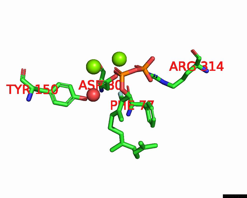
Mono view
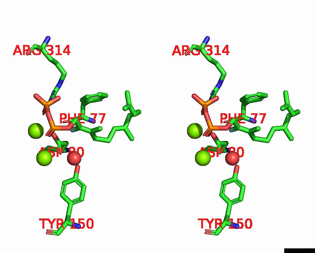
Stereo pair view
Mono view
Stereo pair view
A full contact list of Fluorine with other atoms in the F binding
site number 1 of Crystal Structure of Pentalenene Synthase Variant F76A Complexed with 2-Fluorofarnesyl Diphosphate within 5.0Å range:
|
Reference:
R.P.Kumar,
J.O.Matos,
B.Y.Black,
W.H.Ellenburg,
J.Chen,
M.Patterson,
J.A.Gehtman,
D.L.Theobald,
I.J.Krauss,
D.D.Oprian.
Crystal Structure of Caryolan-1-Ol Synthase, A Sesquiterpene Synthase Catalyzing An Initial Anti-Markovnikov Cyclization Reaction. Biochemistry 2024.
ISSN: ISSN 0006-2960
PubMed: 39400323
DOI: 10.1021/ACS.BIOCHEM.4C00547
Page generated: Wed Jul 16 10:40:14 2025
ISSN: ISSN 0006-2960
PubMed: 39400323
DOI: 10.1021/ACS.BIOCHEM.4C00547
Last articles
Fe in 2CW2Fe in 2CKF
Fe in 2CSG
Fe in 2CPP
Fe in 2CPO
Fe in 2CLB
Fe in 2CP4
Fe in 2CN4
Fe in 2CMN
Fe in 2CMM