Fluorine »
PDB 9cdl-9f2h »
9cjp »
Fluorine in PDB 9cjp: X-Ray Crystal Structure of Sars-Cov-2 Main Protease Quadruple Mutants in Complex with Nirmatrelvir
Enzymatic activity of X-Ray Crystal Structure of Sars-Cov-2 Main Protease Quadruple Mutants in Complex with Nirmatrelvir
All present enzymatic activity of X-Ray Crystal Structure of Sars-Cov-2 Main Protease Quadruple Mutants in Complex with Nirmatrelvir:
3.4.22.69;
3.4.22.69;
Protein crystallography data
The structure of X-Ray Crystal Structure of Sars-Cov-2 Main Protease Quadruple Mutants in Complex with Nirmatrelvir, PDB code: 9cjp
was solved by
M.A.Esler,
K.Shi,
R.S.Harris,
H.Aihara,
with X-Ray Crystallography technique. A brief refinement statistics is given in the table below:
| Resolution Low / High (Å) | 52.83 / 1.71 |
| Space group | P 1 21 1 |
| Cell size a, b, c (Å), α, β, γ (°) | 48.095, 105.665, 53.1, 90, 103.61, 90 |
| R / Rfree (%) | 18 / 22.6 |
Other elements in 9cjp:
The structure of X-Ray Crystal Structure of Sars-Cov-2 Main Protease Quadruple Mutants in Complex with Nirmatrelvir also contains other interesting chemical elements:
| Bromine | (Br) | 3 atoms |
Fluorine Binding Sites:
The binding sites of Fluorine atom in the X-Ray Crystal Structure of Sars-Cov-2 Main Protease Quadruple Mutants in Complex with Nirmatrelvir
(pdb code 9cjp). This binding sites where shown within
5.0 Angstroms radius around Fluorine atom.
In total 6 binding sites of Fluorine where determined in the X-Ray Crystal Structure of Sars-Cov-2 Main Protease Quadruple Mutants in Complex with Nirmatrelvir, PDB code: 9cjp:
Jump to Fluorine binding site number: 1; 2; 3; 4; 5; 6;
In total 6 binding sites of Fluorine where determined in the X-Ray Crystal Structure of Sars-Cov-2 Main Protease Quadruple Mutants in Complex with Nirmatrelvir, PDB code: 9cjp:
Jump to Fluorine binding site number: 1; 2; 3; 4; 5; 6;
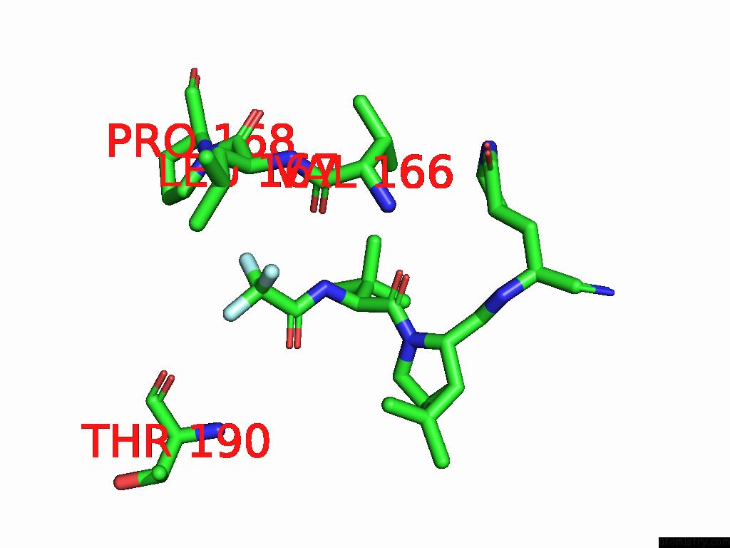
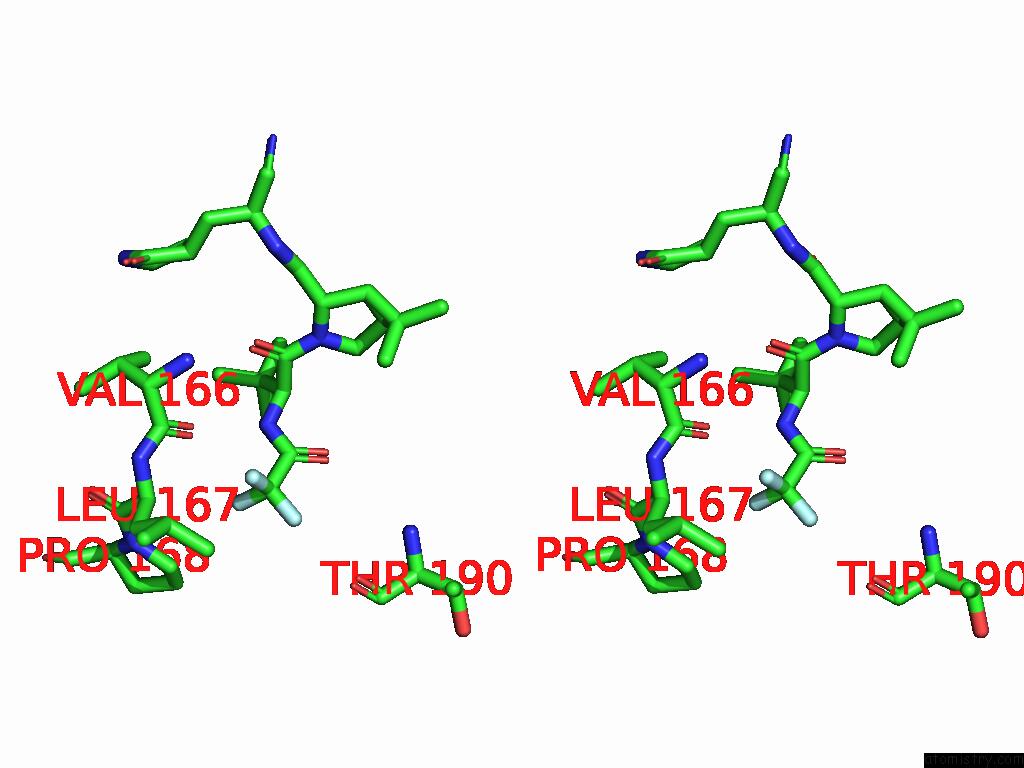
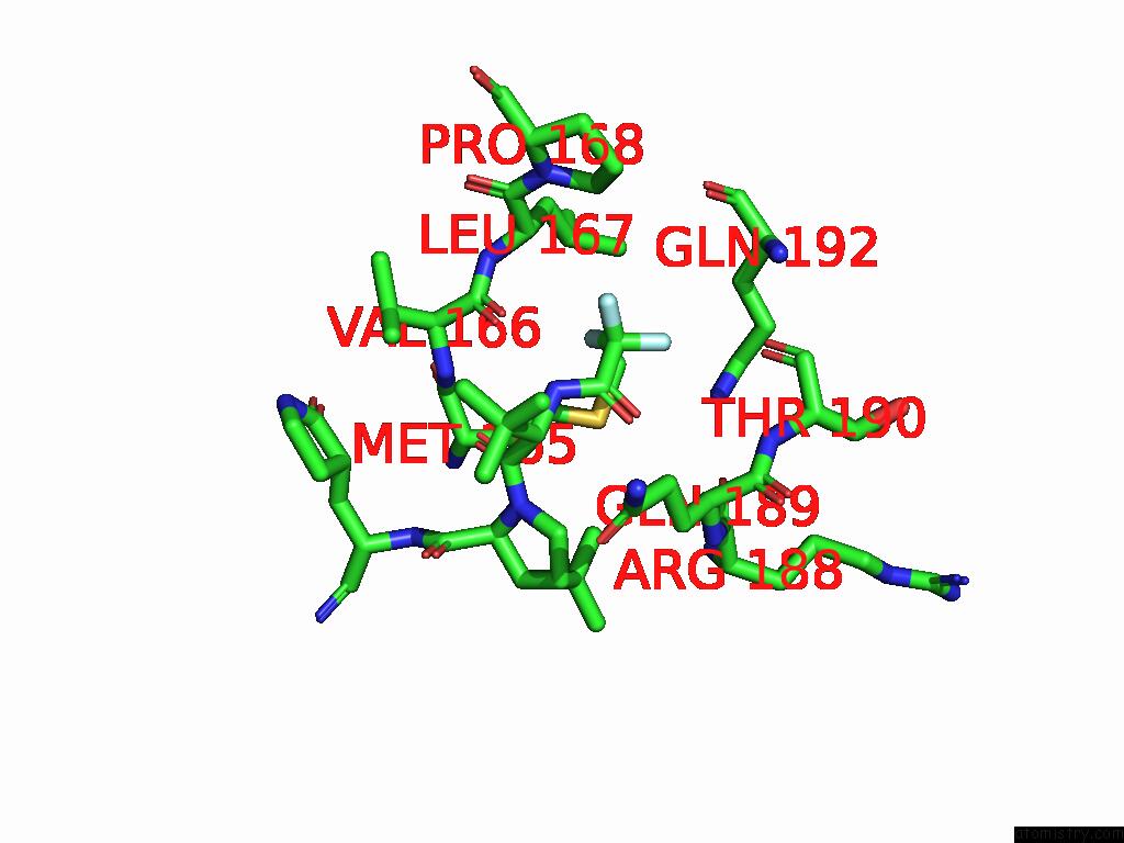
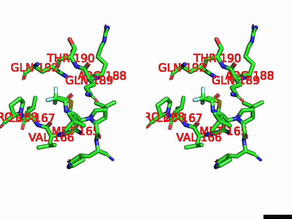
Fluorine binding site 1 out of 6 in 9cjp
Go back to
Fluorine binding site 1 out
of 6 in the X-Ray Crystal Structure of Sars-Cov-2 Main Protease Quadruple Mutants in Complex with Nirmatrelvir
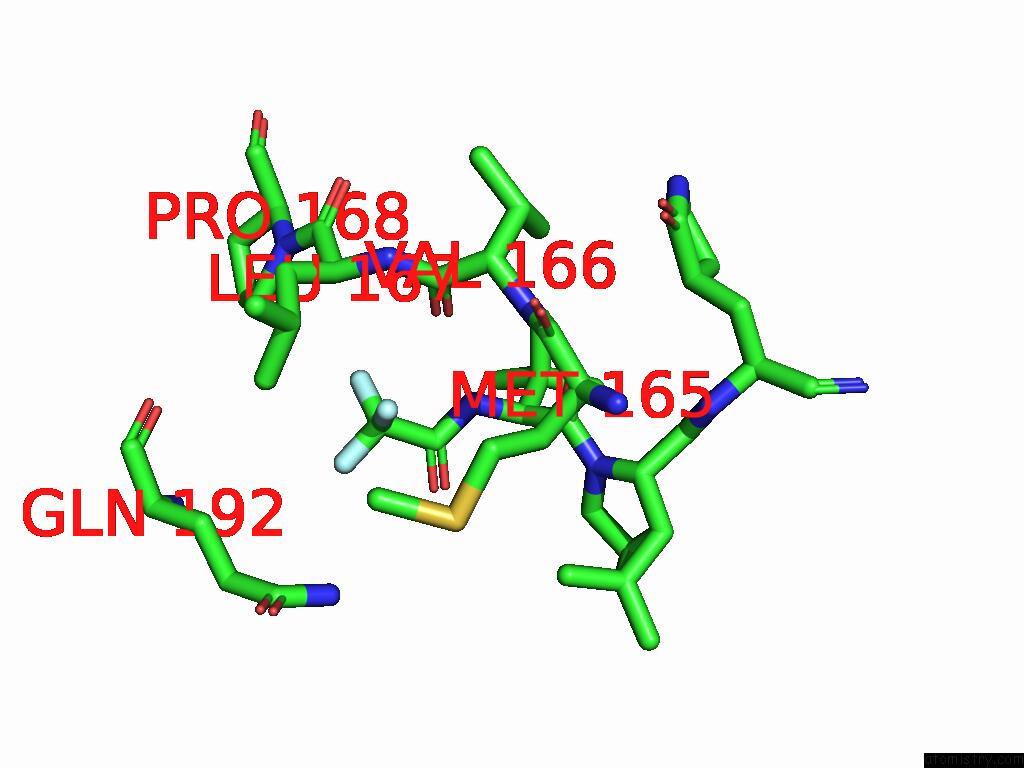
Mono view
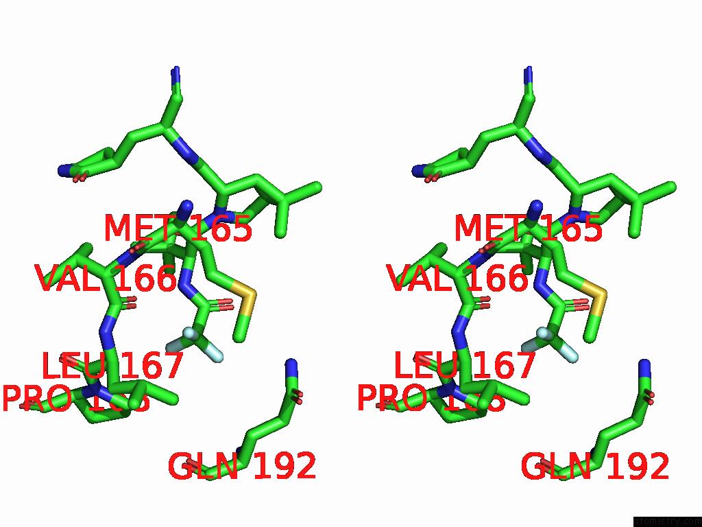
Stereo pair view
Mono view
Stereo pair view
A full contact list of Fluorine with other atoms in the F binding
site number 1 of X-Ray Crystal Structure of Sars-Cov-2 Main Protease Quadruple Mutants in Complex with Nirmatrelvir within 5.0Å range:
|
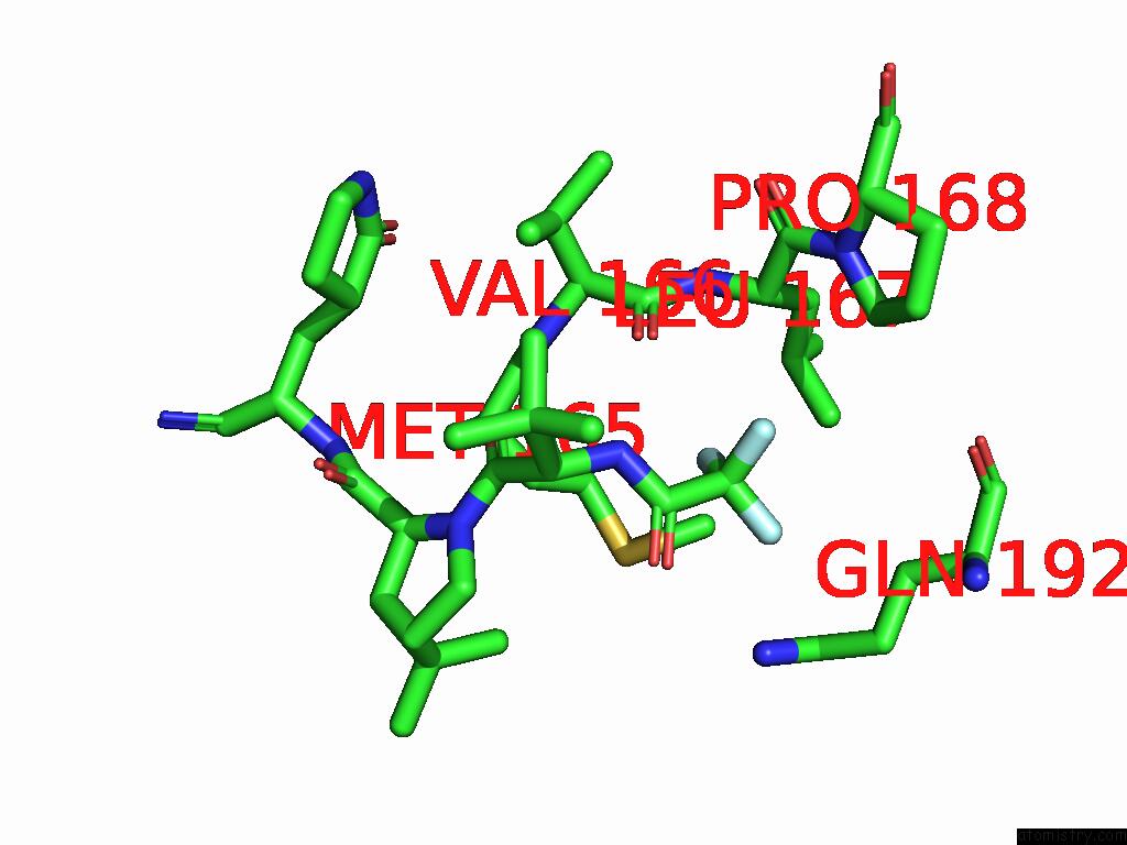
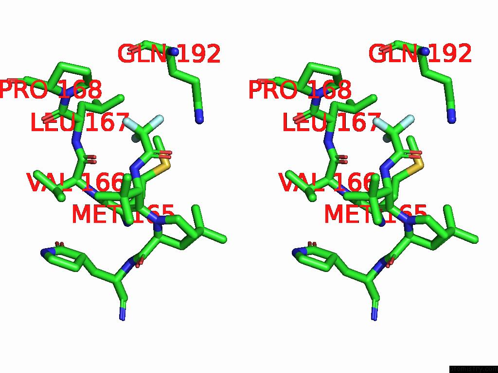
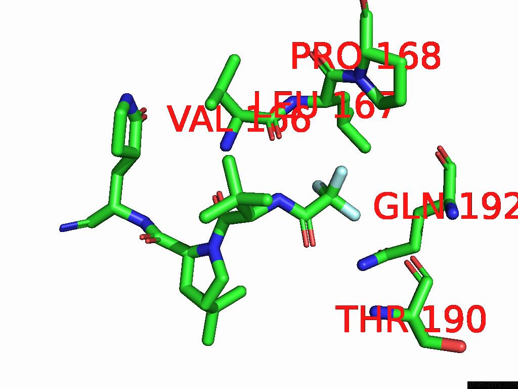
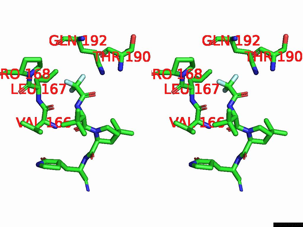
Fluorine binding site 2 out of 6 in 9cjp
Go back to
Fluorine binding site 2 out
of 6 in the X-Ray Crystal Structure of Sars-Cov-2 Main Protease Quadruple Mutants in Complex with Nirmatrelvir
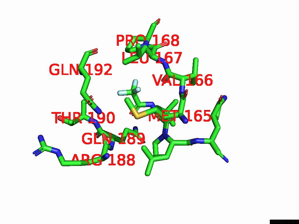
Mono view
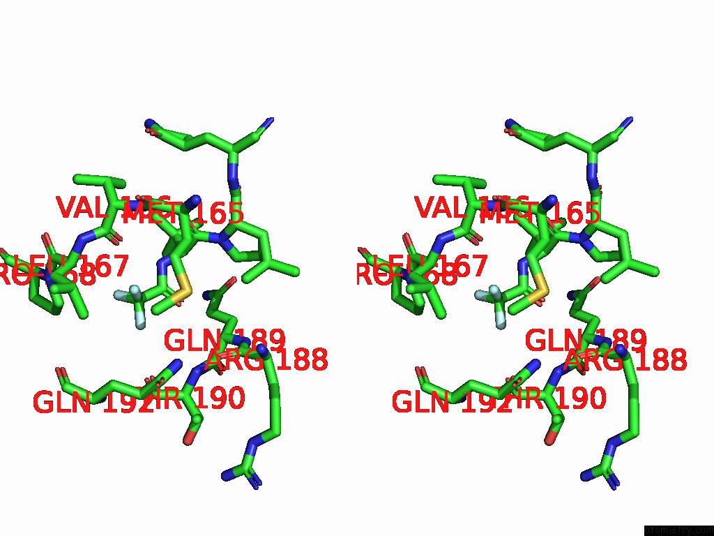
Stereo pair view
Mono view
Stereo pair view
A full contact list of Fluorine with other atoms in the F binding
site number 2 of X-Ray Crystal Structure of Sars-Cov-2 Main Protease Quadruple Mutants in Complex with Nirmatrelvir within 5.0Å range:
|
Fluorine binding site 3 out of 6 in 9cjp
Go back to
Fluorine binding site 3 out
of 6 in the X-Ray Crystal Structure of Sars-Cov-2 Main Protease Quadruple Mutants in Complex with Nirmatrelvir
Mono view
Stereo pair view
Mono view
Stereo pair view
A full contact list of Fluorine with other atoms in the F binding
site number 3 of X-Ray Crystal Structure of Sars-Cov-2 Main Protease Quadruple Mutants in Complex with Nirmatrelvir within 5.0Å range:
|
Fluorine binding site 4 out of 6 in 9cjp
Go back to
Fluorine binding site 4 out
of 6 in the X-Ray Crystal Structure of Sars-Cov-2 Main Protease Quadruple Mutants in Complex with Nirmatrelvir
Mono view
Stereo pair view
Mono view
Stereo pair view
A full contact list of Fluorine with other atoms in the F binding
site number 4 of X-Ray Crystal Structure of Sars-Cov-2 Main Protease Quadruple Mutants in Complex with Nirmatrelvir within 5.0Å range:
|
Fluorine binding site 5 out of 6 in 9cjp
Go back to
Fluorine binding site 5 out
of 6 in the X-Ray Crystal Structure of Sars-Cov-2 Main Protease Quadruple Mutants in Complex with Nirmatrelvir
Mono view
Stereo pair view
Mono view
Stereo pair view
A full contact list of Fluorine with other atoms in the F binding
site number 5 of X-Ray Crystal Structure of Sars-Cov-2 Main Protease Quadruple Mutants in Complex with Nirmatrelvir within 5.0Å range:
|
Fluorine binding site 6 out of 6 in 9cjp
Go back to
Fluorine binding site 6 out
of 6 in the X-Ray Crystal Structure of Sars-Cov-2 Main Protease Quadruple Mutants in Complex with Nirmatrelvir
Mono view
Stereo pair view
Mono view
Stereo pair view
A full contact list of Fluorine with other atoms in the F binding
site number 6 of X-Ray Crystal Structure of Sars-Cov-2 Main Protease Quadruple Mutants in Complex with Nirmatrelvir within 5.0Å range:
|
Reference:
M.A.Esler,
K.Shi,
R.S.Harris,
H.Aihara.
X-Ray Crystal Structure of Sars-Cov-2 Main Protease Quadruple Mutants in Complex with Nirmatrelvir To Be Published.
Page generated: Wed Jul 16 10:42:51 2025
Last articles
Fe in 1V4UFe in 1UZR
Fe in 1V0H
Fe in 1V07
Fe in 1UWV
Fe in 1UX9
Fe in 1UZW
Fe in 1UYU
Fe in 1UWM
Fe in 1UX8