Fluorine »
PDB 9f2l-9msq »
9ix2 »
Fluorine in PDB 9ix2: Crystal Structure of the Mouse RIP3 Kinase Domain in Complexed with Tak-632
Enzymatic activity of Crystal Structure of the Mouse RIP3 Kinase Domain in Complexed with Tak-632
All present enzymatic activity of Crystal Structure of the Mouse RIP3 Kinase Domain in Complexed with Tak-632:
2.7.11.1;
2.7.11.1;
Protein crystallography data
The structure of Crystal Structure of the Mouse RIP3 Kinase Domain in Complexed with Tak-632, PDB code: 9ix2
was solved by
H.Xie,
H.X.Su,
M.J.Li,
Y.C.Xu,
with X-Ray Crystallography technique. A brief refinement statistics is given in the table below:
| Resolution Low / High (Å) | 38.89 / 2.39 |
| Space group | C 1 2 1 |
| Cell size a, b, c (Å), α, β, γ (°) | 146.771, 54.87, 102.724, 90, 130.78, 90 |
| R / Rfree (%) | 22.8 / 27.6 |
Fluorine Binding Sites:
The binding sites of Fluorine atom in the Crystal Structure of the Mouse RIP3 Kinase Domain in Complexed with Tak-632
(pdb code 9ix2). This binding sites where shown within
5.0 Angstroms radius around Fluorine atom.
In total 4 binding sites of Fluorine where determined in the Crystal Structure of the Mouse RIP3 Kinase Domain in Complexed with Tak-632, PDB code: 9ix2:
Jump to Fluorine binding site number: 1; 2; 3; 4;
In total 4 binding sites of Fluorine where determined in the Crystal Structure of the Mouse RIP3 Kinase Domain in Complexed with Tak-632, PDB code: 9ix2:
Jump to Fluorine binding site number: 1; 2; 3; 4;
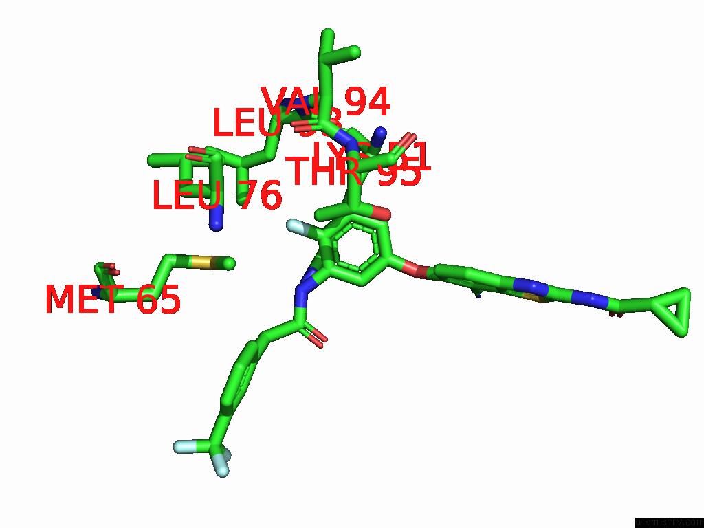
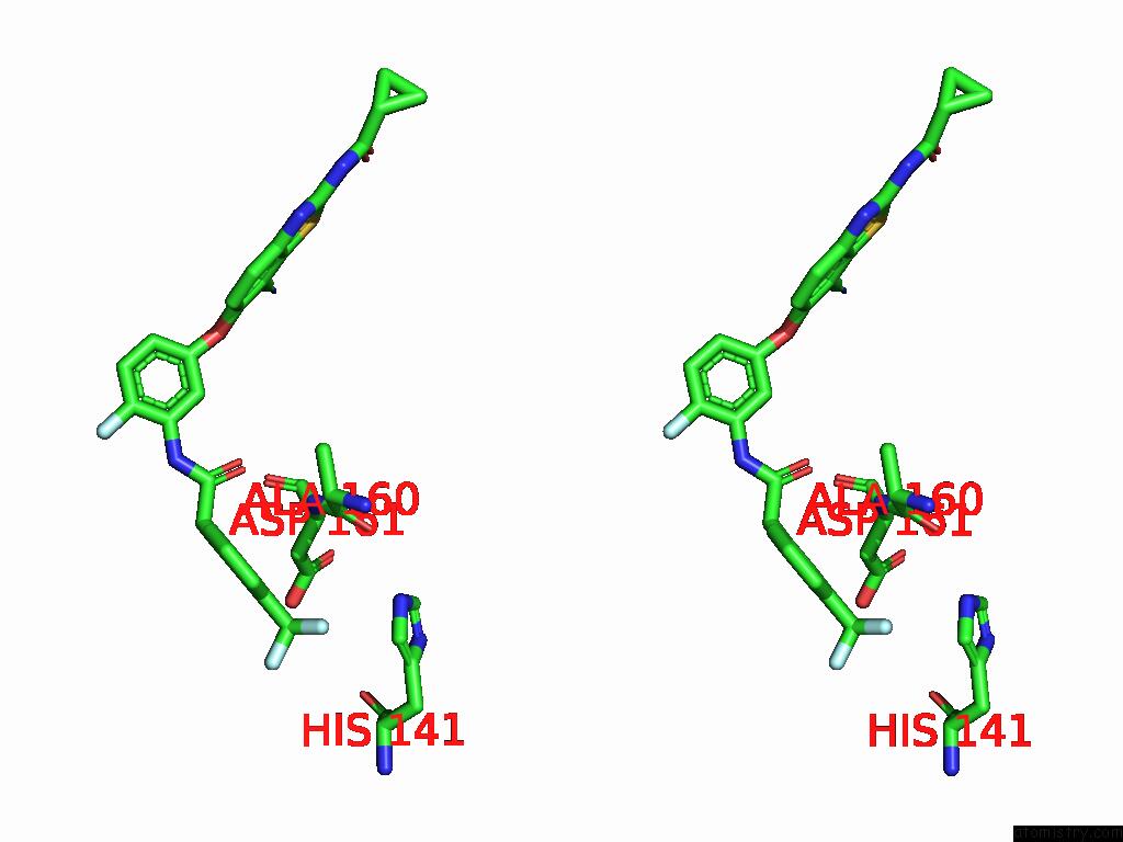
Fluorine binding site 1 out of 4 in 9ix2
Go back to
Fluorine binding site 1 out
of 4 in the Crystal Structure of the Mouse RIP3 Kinase Domain in Complexed with Tak-632
Mono view
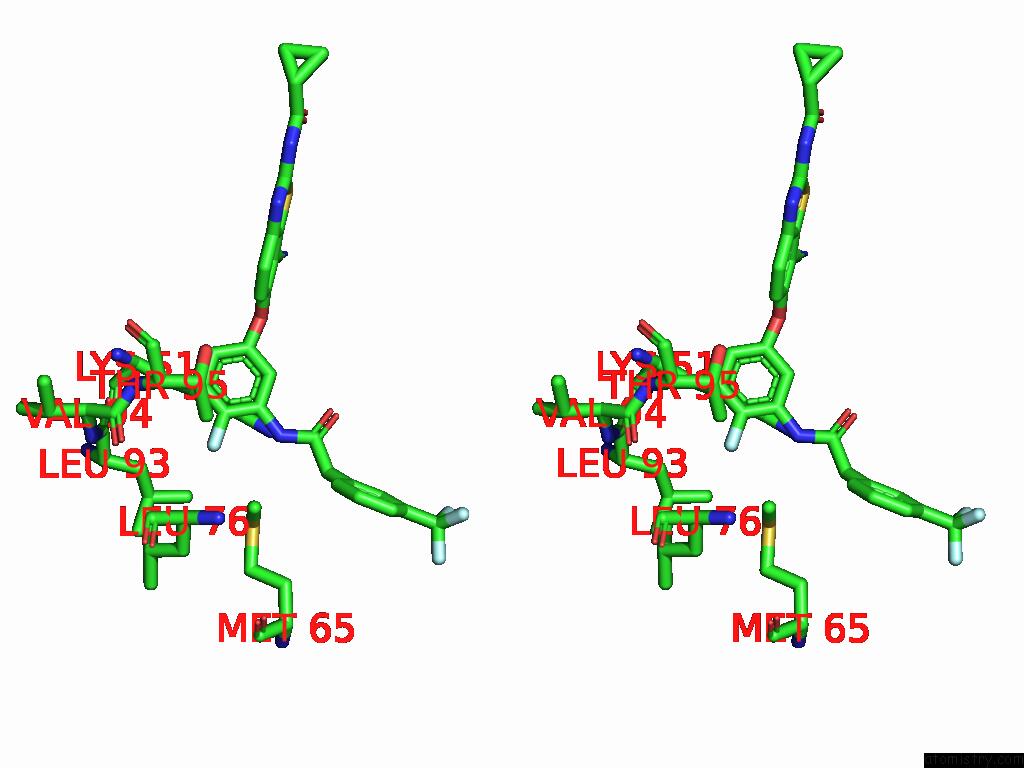
Stereo pair view
Mono view
Stereo pair view
A full contact list of Fluorine with other atoms in the F binding
site number 1 of Crystal Structure of the Mouse RIP3 Kinase Domain in Complexed with Tak-632 within 5.0Å range:
|
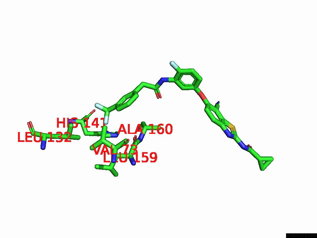
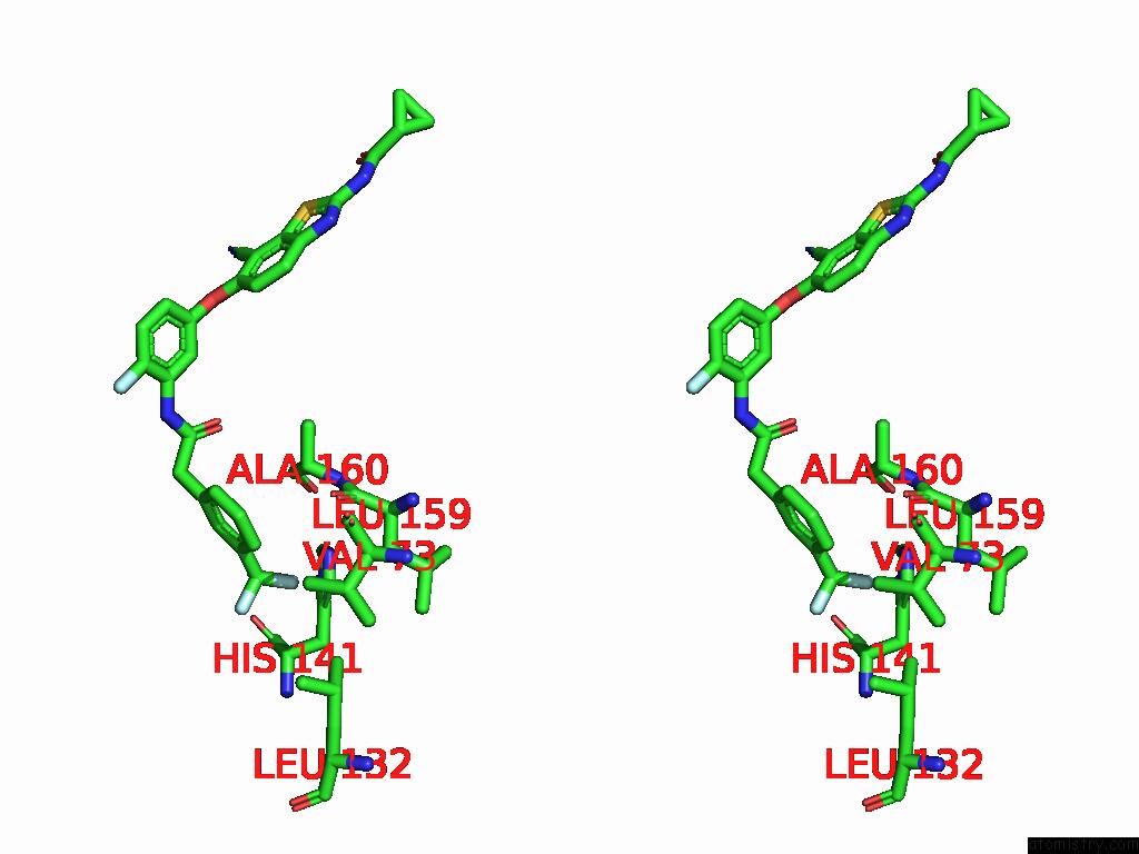
Fluorine binding site 2 out of 4 in 9ix2
Go back to
Fluorine binding site 2 out
of 4 in the Crystal Structure of the Mouse RIP3 Kinase Domain in Complexed with Tak-632
Mono view
Stereo pair view
Mono view
Stereo pair view
A full contact list of Fluorine with other atoms in the F binding
site number 2 of Crystal Structure of the Mouse RIP3 Kinase Domain in Complexed with Tak-632 within 5.0Å range:
|
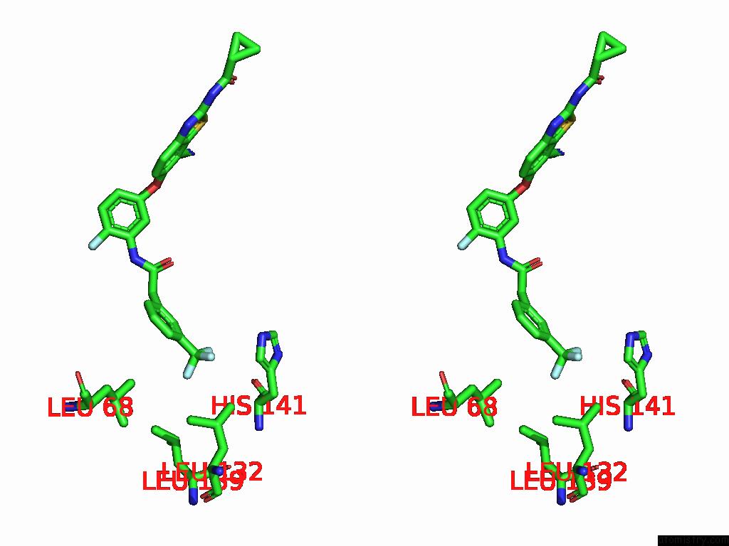
Fluorine binding site 3 out of 4 in 9ix2
Go back to
Fluorine binding site 3 out
of 4 in the Crystal Structure of the Mouse RIP3 Kinase Domain in Complexed with Tak-632

Mono view
Stereo pair view
Mono view
Stereo pair view
A full contact list of Fluorine with other atoms in the F binding
site number 3 of Crystal Structure of the Mouse RIP3 Kinase Domain in Complexed with Tak-632 within 5.0Å range:
|
Fluorine binding site 4 out of 4 in 9ix2
Go back to
Fluorine binding site 4 out
of 4 in the Crystal Structure of the Mouse RIP3 Kinase Domain in Complexed with Tak-632
Mono view
Stereo pair view
Mono view
Stereo pair view
A full contact list of Fluorine with other atoms in the F binding
site number 4 of Crystal Structure of the Mouse RIP3 Kinase Domain in Complexed with Tak-632 within 5.0Å range:
|
Reference:
H.Xie,
H.X.Su,
M.J.Li,
Y.C.Xu.
Structure-Based Design of Potent and Selective Inhibitors Targeting RIPK3 For Eliminating on-Target Toxicity in Vitro Nat Commun 2025.
ISSN: ESSN 2041-1723
Page generated: Wed Jul 16 11:41:35 2025
ISSN: ESSN 2041-1723
Last articles
Fe in 1JWKFe in 1JWJ
Fe in 1JW8
Fe in 1JRZ
Fe in 1JW1
Fe in 1JRP
Fe in 1JRY
Fe in 1JRX
Fe in 1JRO
Fe in 1JNZ