Fluorine »
PDB 5tbo-5tvj »
5tm1 »
Fluorine in PDB 5tm1: Crystal Structure of the Er-Alpha Ligand-Binding Domain (Y537S) in Complex with 2,5-Bis(2-Fluoro-4-Hydroxyphenyl)Thiophene 1-Oxide
Protein crystallography data
The structure of Crystal Structure of the Er-Alpha Ligand-Binding Domain (Y537S) in Complex with 2,5-Bis(2-Fluoro-4-Hydroxyphenyl)Thiophene 1-Oxide, PDB code: 5tm1
was solved by
J.C.Nwachukwu,
N.J.Wright,
S.Srinivasan,
N.E.Bruno,
J.Nowak,
D.J.Kojetin,
O.Elemento,
J.A.Katzenellenbogen,
K.W.Nettles,
with X-Ray Crystallography technique. A brief refinement statistics is given in the table below:
| Resolution Low / High (Å) | 47.27 / 2.23 |
| Space group | P 1 21 1 |
| Cell size a, b, c (Å), α, β, γ (°) | 55.980, 83.040, 59.000, 90.00, 110.84, 90.00 |
| R / Rfree (%) | 19.8 / 23.6 |
Fluorine Binding Sites:
The binding sites of Fluorine atom in the Crystal Structure of the Er-Alpha Ligand-Binding Domain (Y537S) in Complex with 2,5-Bis(2-Fluoro-4-Hydroxyphenyl)Thiophene 1-Oxide
(pdb code 5tm1). This binding sites where shown within
5.0 Angstroms radius around Fluorine atom.
In total 4 binding sites of Fluorine where determined in the Crystal Structure of the Er-Alpha Ligand-Binding Domain (Y537S) in Complex with 2,5-Bis(2-Fluoro-4-Hydroxyphenyl)Thiophene 1-Oxide, PDB code: 5tm1:
Jump to Fluorine binding site number: 1; 2; 3; 4;
In total 4 binding sites of Fluorine where determined in the Crystal Structure of the Er-Alpha Ligand-Binding Domain (Y537S) in Complex with 2,5-Bis(2-Fluoro-4-Hydroxyphenyl)Thiophene 1-Oxide, PDB code: 5tm1:
Jump to Fluorine binding site number: 1; 2; 3; 4;
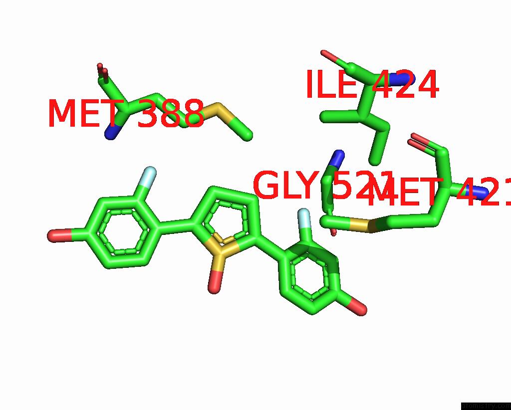
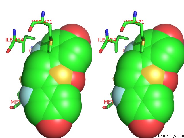
Fluorine binding site 1 out of 4 in 5tm1
Go back to
Fluorine binding site 1 out
of 4 in the Crystal Structure of the Er-Alpha Ligand-Binding Domain (Y537S) in Complex with 2,5-Bis(2-Fluoro-4-Hydroxyphenyl)Thiophene 1-Oxide
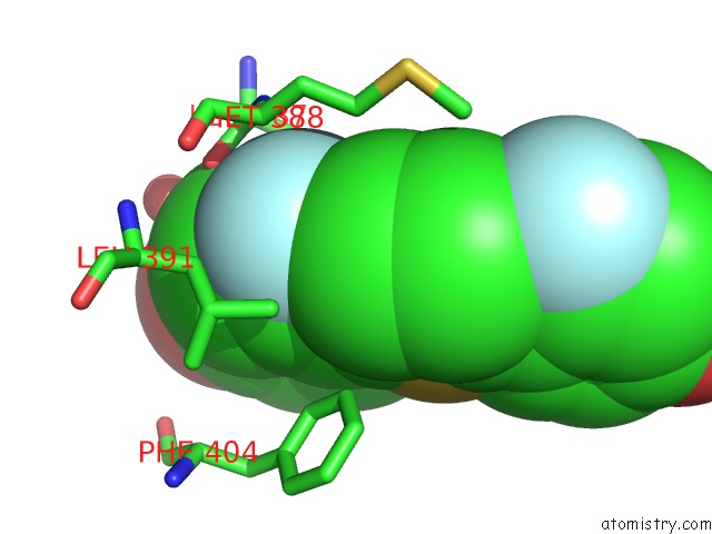
Mono view
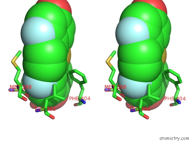
Stereo pair view
Mono view
Stereo pair view
A full contact list of Fluorine with other atoms in the F binding
site number 1 of Crystal Structure of the Er-Alpha Ligand-Binding Domain (Y537S) in Complex with 2,5-Bis(2-Fluoro-4-Hydroxyphenyl)Thiophene 1-Oxide within 5.0Å range:
|
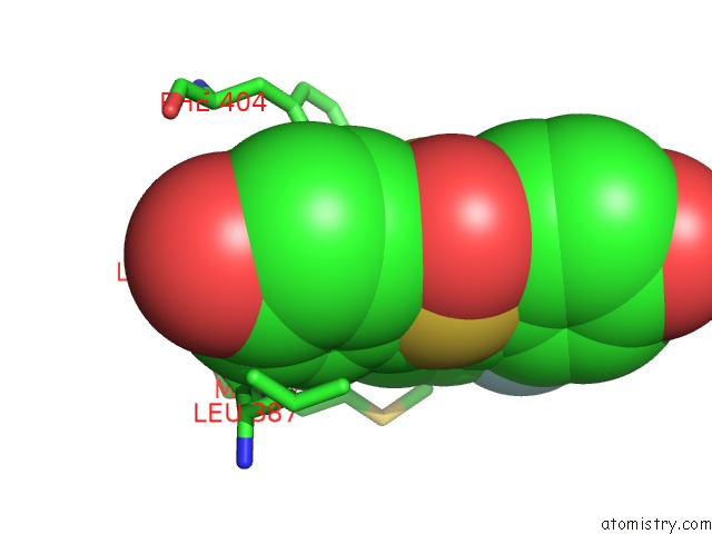
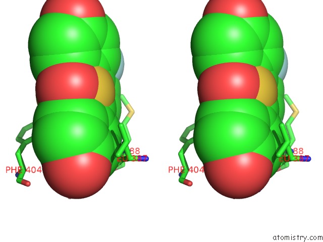
Fluorine binding site 2 out of 4 in 5tm1
Go back to
Fluorine binding site 2 out
of 4 in the Crystal Structure of the Er-Alpha Ligand-Binding Domain (Y537S) in Complex with 2,5-Bis(2-Fluoro-4-Hydroxyphenyl)Thiophene 1-Oxide
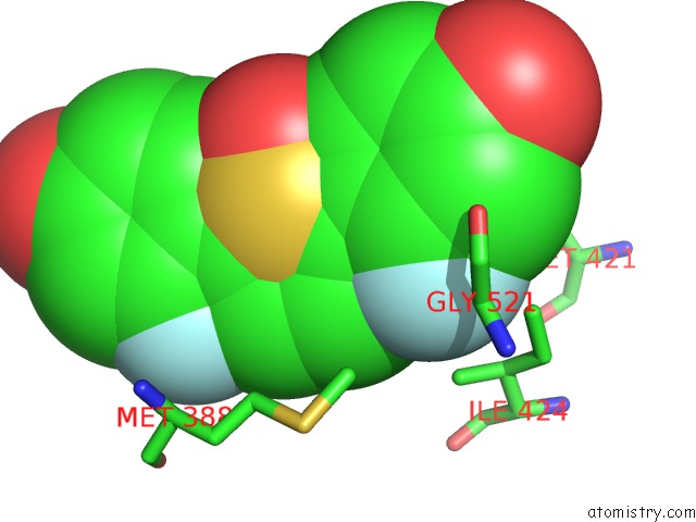
Mono view
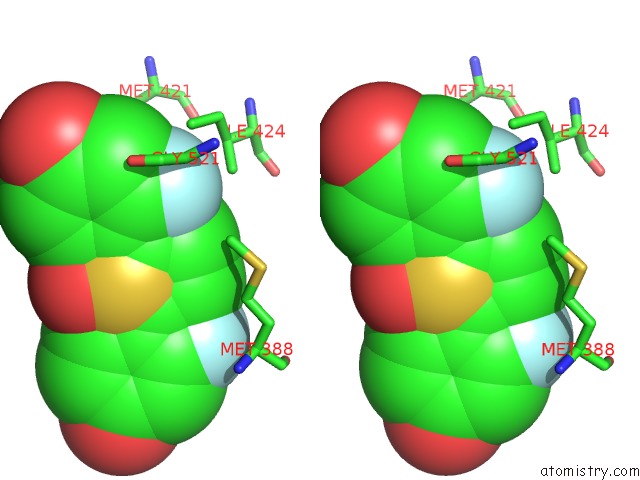
Stereo pair view
Mono view
Stereo pair view
A full contact list of Fluorine with other atoms in the F binding
site number 2 of Crystal Structure of the Er-Alpha Ligand-Binding Domain (Y537S) in Complex with 2,5-Bis(2-Fluoro-4-Hydroxyphenyl)Thiophene 1-Oxide within 5.0Å range:
|
Fluorine binding site 3 out of 4 in 5tm1
Go back to
Fluorine binding site 3 out
of 4 in the Crystal Structure of the Er-Alpha Ligand-Binding Domain (Y537S) in Complex with 2,5-Bis(2-Fluoro-4-Hydroxyphenyl)Thiophene 1-Oxide
Mono view
Stereo pair view
Mono view
Stereo pair view
A full contact list of Fluorine with other atoms in the F binding
site number 3 of Crystal Structure of the Er-Alpha Ligand-Binding Domain (Y537S) in Complex with 2,5-Bis(2-Fluoro-4-Hydroxyphenyl)Thiophene 1-Oxide within 5.0Å range:
|
Fluorine binding site 4 out of 4 in 5tm1
Go back to
Fluorine binding site 4 out
of 4 in the Crystal Structure of the Er-Alpha Ligand-Binding Domain (Y537S) in Complex with 2,5-Bis(2-Fluoro-4-Hydroxyphenyl)Thiophene 1-Oxide
Mono view
Stereo pair view
Mono view
Stereo pair view
A full contact list of Fluorine with other atoms in the F binding
site number 4 of Crystal Structure of the Er-Alpha Ligand-Binding Domain (Y537S) in Complex with 2,5-Bis(2-Fluoro-4-Hydroxyphenyl)Thiophene 1-Oxide within 5.0Å range:
|
Reference:
J.C.Nwachukwu,
S.Srinivasan,
N.E.Bruno,
J.Nowak,
N.J.Wright,
F.Minutolo,
E.S.Rangarajan,
T.Izard,
X.Q.Yao,
B.J.Grant,
D.J.Kojetin,
O.Elemento,
J.A.Katzenellenbogen,
K.W.Nettles.
Systems Structural Biology Analysis of Ligand Effects on Er Alpha Predicts Cellular Response to Environmental Estrogens and Anti-Hormone Therapies. Cell Chem Biol V. 24 35 2017.
ISSN: ESSN 2451-9456
PubMed: 28042045
DOI: 10.1016/J.CHEMBIOL.2016.11.014
Page generated: Tue Jul 15 07:59:13 2025
ISSN: ESSN 2451-9456
PubMed: 28042045
DOI: 10.1016/J.CHEMBIOL.2016.11.014
Last articles
Mg in 1IZLMg in 1JBZ
Mg in 1JBW
Mg in 1JBV
Mg in 1JBK
Mg in 1JAX
Mg in 1JAH
Mg in 1J97
Mg in 1J9J
Mg in 1J8L