Fluorine »
PDB 1xz3-1zzr »
1z34 »
Fluorine in PDB 1z34: Crystal Structure of Trichomonas Vaginalis Purine Nucleoside Phosphorylase Complexed with 2-Fluoro-2'-Deoxyadenosine
Enzymatic activity of Crystal Structure of Trichomonas Vaginalis Purine Nucleoside Phosphorylase Complexed with 2-Fluoro-2'-Deoxyadenosine
All present enzymatic activity of Crystal Structure of Trichomonas Vaginalis Purine Nucleoside Phosphorylase Complexed with 2-Fluoro-2'-Deoxyadenosine:
2.4.2.1;
2.4.2.1;
Protein crystallography data
The structure of Crystal Structure of Trichomonas Vaginalis Purine Nucleoside Phosphorylase Complexed with 2-Fluoro-2'-Deoxyadenosine, PDB code: 1z34
was solved by
Y.Zhang,
W.H.Wang,
S.W.Wu,
C.C.Wang,
S.E.Ealick,
with X-Ray Crystallography technique. A brief refinement statistics is given in the table below:
| Resolution Low / High (Å) | 37.94 / 2.40 |
| Space group | P 41 3 2 |
| Cell size a, b, c (Å), α, β, γ (°) | 136.800, 136.800, 136.800, 90.00, 90.00, 90.00 |
| R / Rfree (%) | 20.4 / 24 |
Fluorine Binding Sites:
The binding sites of Fluorine atom in the Crystal Structure of Trichomonas Vaginalis Purine Nucleoside Phosphorylase Complexed with 2-Fluoro-2'-Deoxyadenosine
(pdb code 1z34). This binding sites where shown within
5.0 Angstroms radius around Fluorine atom.
In total only one binding site of Fluorine was determined in the Crystal Structure of Trichomonas Vaginalis Purine Nucleoside Phosphorylase Complexed with 2-Fluoro-2'-Deoxyadenosine, PDB code: 1z34:
In total only one binding site of Fluorine was determined in the Crystal Structure of Trichomonas Vaginalis Purine Nucleoside Phosphorylase Complexed with 2-Fluoro-2'-Deoxyadenosine, PDB code: 1z34:
Fluorine binding site 1 out of 1 in 1z34
Go back to
Fluorine binding site 1 out
of 1 in the Crystal Structure of Trichomonas Vaginalis Purine Nucleoside Phosphorylase Complexed with 2-Fluoro-2'-Deoxyadenosine
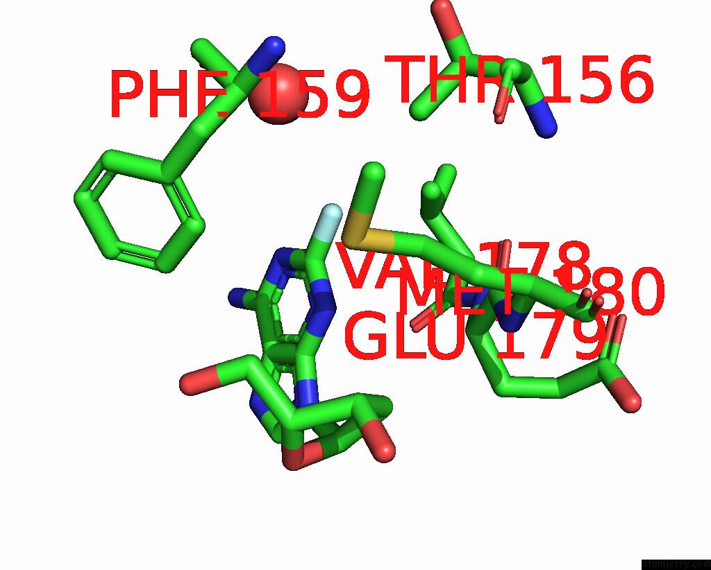
Mono view
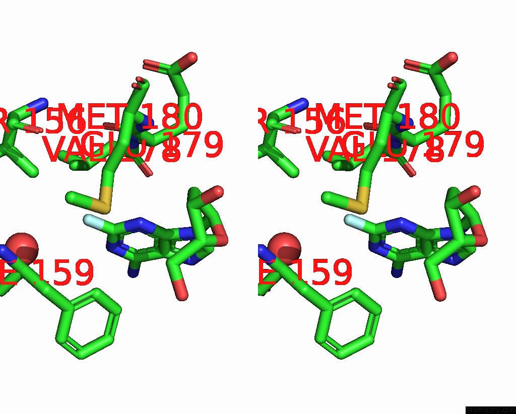
Stereo pair view
Mono view
Stereo pair view
A full contact list of Fluorine with other atoms in the F binding
site number 1 of Crystal Structure of Trichomonas Vaginalis Purine Nucleoside Phosphorylase Complexed with 2-Fluoro-2'-Deoxyadenosine within 5.0Å range:
|
Reference:
Y.Zang,
W.H.Wang,
S.W.Wu,
S.E.Ealick,
C.C.Wang.
Identification of A Subversive Substrate of Trichomonas Vaginalis Purine Nucleoside Phosphorylase and the Crystal Structure of the Enzyme-Substrate Complex. J.Biol.Chem. V. 280 22318 2005.
ISSN: ISSN 0021-9258
PubMed: 15817485
DOI: 10.1074/JBC.M501843200
Page generated: Mon Jul 14 12:28:56 2025
ISSN: ISSN 0021-9258
PubMed: 15817485
DOI: 10.1074/JBC.M501843200
Last articles
F in 7LD3F in 7LCR
F in 7LCM
F in 7LCO
F in 7LCK
F in 7LCJ
F in 7LCI
F in 7L9Y
F in 7LCD
F in 7LAY