Atomistry » Fluorine » PDB 3vrs-3wyk » 3w0y
Atomistry »
  Fluorine »
    PDB 3vrs-3wyk »
      3w0y »

Fluorine in PDB 3w0y: Crystal Structure Analysis of Vitamin D Receptor

Protein crystallography data

The structure of Crystal Structure Analysis of Vitamin D Receptor, PDB code: 3w0y was solved by S.Itoh, S.Iijima, with X-Ray Crystallography technique. A brief refinement statistics is given in the table below:

Resolution Low / High (Å) 33.87 / 1.98
Space group P 21 21 21
Cell size a, b, c (Å), α, β, γ (°) 45.217, 51.124, 132.664, 90.00, 90.00, 90.00
R / Rfree (%) 17.4 / 21.8

Fluorine Binding Sites:

The binding sites of Fluorine atom in the Crystal Structure Analysis of Vitamin D Receptor (pdb code 3w0y). This binding sites where shown within 5.0 Angstroms radius around Fluorine atom.
In total 7 binding sites of Fluorine where determined in the Crystal Structure Analysis of Vitamin D Receptor, PDB code: 3w0y:
Jump to Fluorine binding site number: 1; 2; 3; 4; 5; 6; 7;

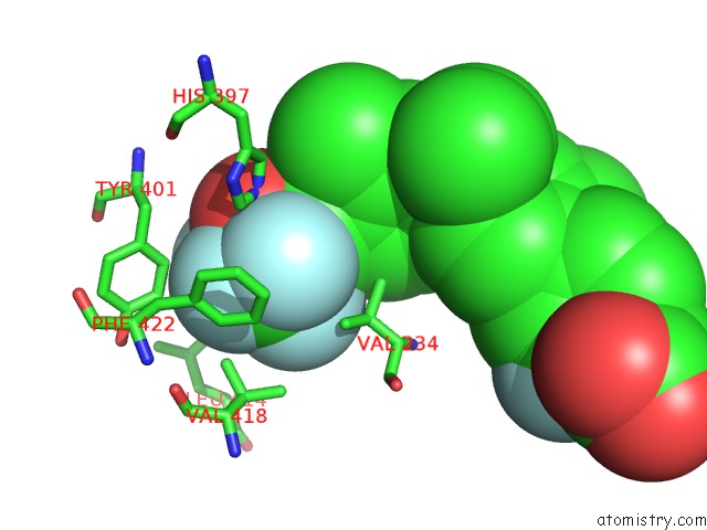
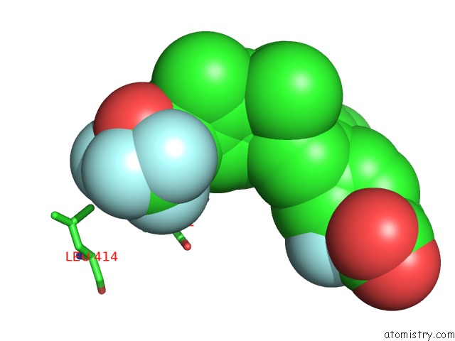
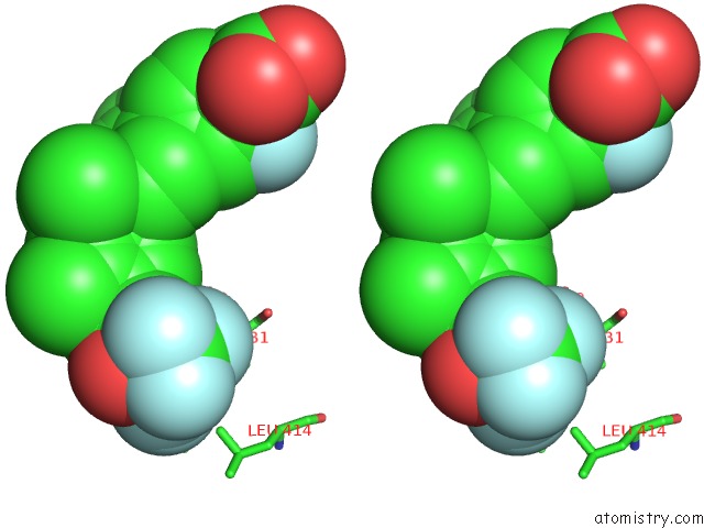
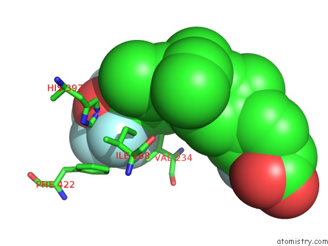
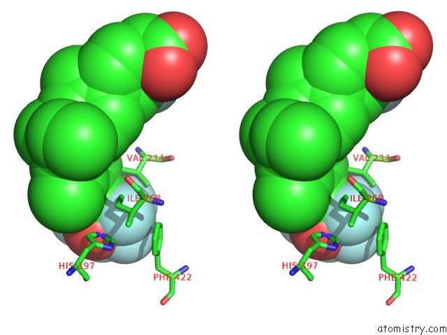
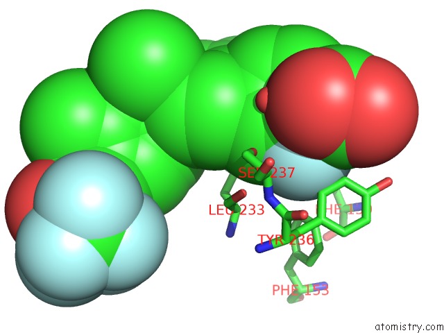
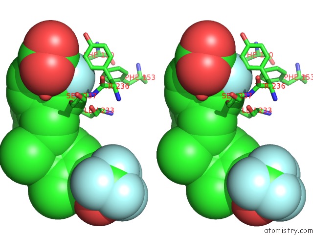
Fluorine binding site 1 out of 7 in 3w0y

Go back to Fluorine Binding Sites List in 3w0y
Fluorine binding site 1 out of 7 in the Crystal Structure Analysis of Vitamin D Receptor


Mono view


Stereo pair view

A full contact list of Fluorine with other atoms in the F binding site number 1 of Crystal Structure Analysis of Vitamin D Receptor within 5.0Å range:
probe atom residue distance (Å) B Occ
A:F501

b:28.0
occ:1.00
F5 A:DS4501 0.0 28.0 1.0
C7 A:DS4501 1.5 26.3 1.0
F6 A:DS4501 2.4 26.5 1.0
F4 A:DS4501 2.5 26.7 1.0
C44 A:DS4501 2.6 26.4 1.0
F2 A:DS4501 2.7 27.5 1.0
O47 A:DS4501 2.8 25.9 1.0
C15 A:DS4501 3.2 26.8 1.0
CD1 A:TYR401 3.2 21.2 1.0
CG1 A:VAL418 3.4 19.5 1.0
CE1 A:TYR401 3.5 21.6 1.0
CD1 A:PHE422 3.7 24.3 1.0
F3 A:DS4501 3.8 27.5 1.0
C43 A:DS4501 3.9 25.5 1.0
CE1 A:PHE422 4.0 23.9 1.0
NE2 A:HIS397 4.1 21.3 1.0
CE1 A:HIS397 4.2 21.1 1.0
CG A:PHE422 4.3 22.3 1.0
CG A:TYR401 4.4 19.7 1.0
F1 A:DS4501 4.5 27.1 1.0
CB A:VAL418 4.7 19.8 1.0
CB A:PHE422 4.7 21.6 1.0
CZ A:TYR401 4.7 27.6 1.0
CD2 A:LEU414 4.7 24.7 1.0
CG2 A:VAL418 4.8 19.2 1.0
CZ A:PHE422 4.9 22.6 1.0
CB A:TYR401 4.9 18.4 1.0
CG1 A:VAL234 5.0 21.3 1.0

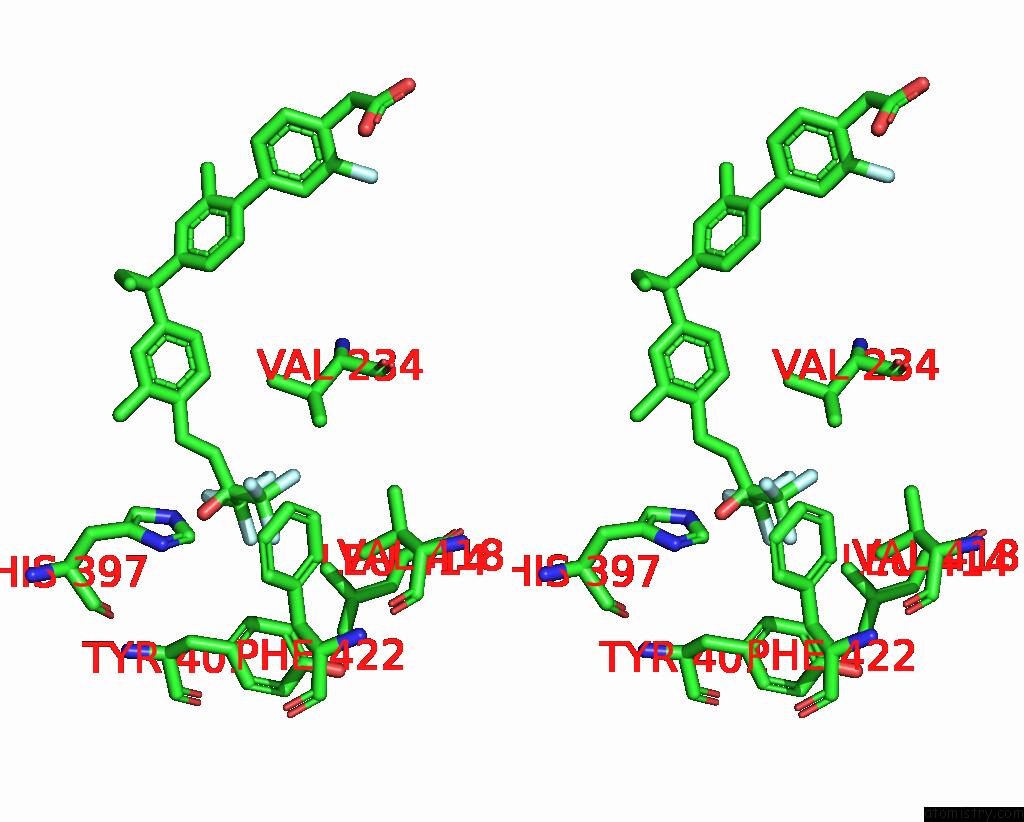
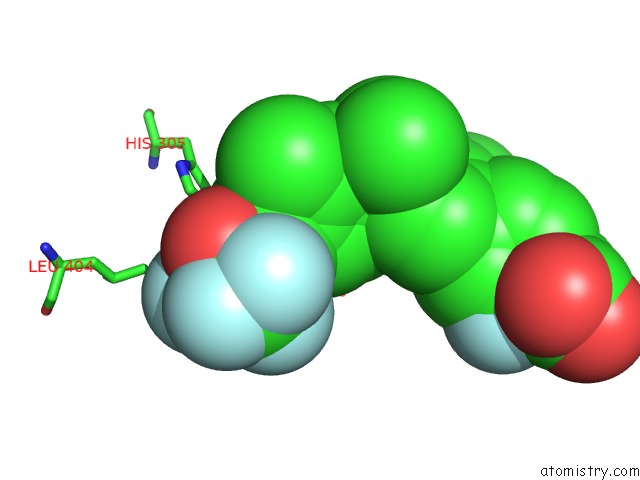
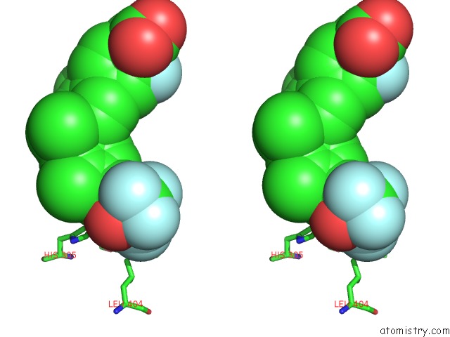
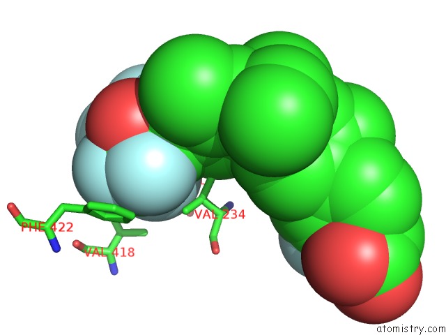
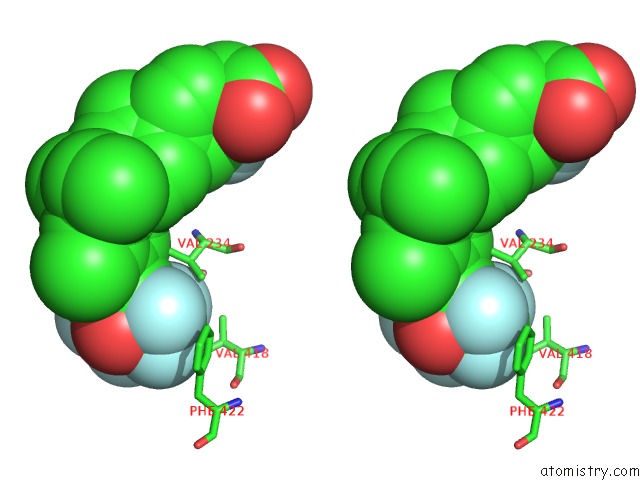
Fluorine binding site 2 out of 7 in 3w0y

Go back to Fluorine Binding Sites List in 3w0y
Fluorine binding site 2 out of 7 in the Crystal Structure Analysis of Vitamin D Receptor


Mono view


Stereo pair view

A full contact list of Fluorine with other atoms in the F binding site number 2 of Crystal Structure Analysis of Vitamin D Receptor within 5.0Å range:
probe atom residue distance (Å) B Occ
A:F501

b:27.5
occ:1.00
F2 A:DS4501 0.0 27.5 1.0
C15 A:DS4501 1.5 26.8 1.0
F1 A:DS4501 2.4 27.1 1.0
F3 A:DS4501 2.4 27.5 1.0
C44 A:DS4501 2.5 26.4 1.0
O47 A:DS4501 2.7 25.9 1.0
F5 A:DS4501 2.7 28.0 1.0
C7 A:DS4501 2.9 26.3 1.0
CD2 A:LEU404 3.3 40.1 1.0
CD1 A:LEU227 3.4 21.1 1.0
F4 A:DS4501 3.4 26.7 1.0
CD2 A:LEU414 3.5 24.7 1.0
C43 A:DS4501 3.9 25.5 1.0
CD1 A:TYR401 4.0 21.2 1.0
CE1 A:TYR401 4.2 21.6 1.0
NE2 A:HIS305 4.3 29.9 1.0
F6 A:DS4501 4.3 26.5 1.0
CG A:LEU404 4.5 35.8 1.0
CG A:LEU227 4.7 21.1 1.0
C8 A:DS4501 4.8 24.1 1.0
CG1 A:VAL418 4.9 19.5 1.0
CG A:LEU414 4.9 23.7 1.0

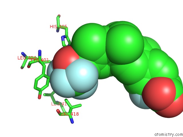
Fluorine binding site 3 out of 7 in 3w0y

Go back to Fluorine Binding Sites List in 3w0y
Fluorine binding site 3 out of 7 in the Crystal Structure Analysis of Vitamin D Receptor


Mono view


Stereo pair view

A full contact list of Fluorine with other atoms in the F binding site number 3 of Crystal Structure Analysis of Vitamin D Receptor within 5.0Å range:
probe atom residue distance (Å) B Occ
A:F501

b:27.5
occ:1.00
F3 A:DS4501 0.0 27.5 1.0
C15 A:DS4501 1.5 26.8 1.0
F2 A:DS4501 2.4 27.5 1.0
F1 A:DS4501 2.5 27.1 1.0
C44 A:DS4501 2.5 26.4 1.0
F4 A:DS4501 2.6 26.7 1.0
C7 A:DS4501 3.0 26.3 1.0
C43 A:DS4501 3.2 25.5 1.0
CA A:ALA231 3.2 17.5 1.0
N A:ALA231 3.3 17.0 1.0
CD1 A:LEU227 3.3 21.1 1.0
CB A:ALA231 3.5 18.3 1.0
O47 A:DS4501 3.6 25.9 1.0
C A:LEU230 3.7 20.8 1.0
F5 A:DS4501 3.8 28.0 1.0
O A:LEU230 4.0 18.9 1.0
C8 A:DS4501 4.1 24.1 1.0
CD2 A:LEU414 4.3 24.7 1.0
O A:LEU227 4.3 18.4 1.0
CB A:LEU230 4.3 20.0 1.0
F6 A:DS4501 4.4 26.5 1.0
CA A:LEU230 4.6 19.3 1.0
CG A:LEU227 4.6 21.1 1.0
C A:ALA231 4.7 20.8 1.0
C29 A:DS4501 4.9 18.5 1.0
C28 A:DS4501 4.9 20.0 1.0

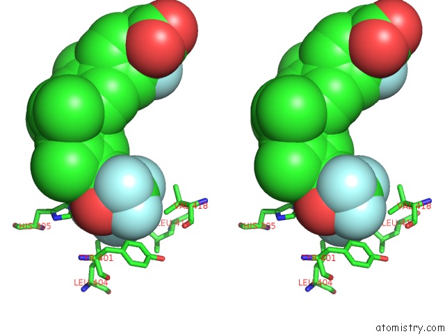
Fluorine binding site 4 out of 7 in 3w0y

Go back to Fluorine Binding Sites List in 3w0y
Fluorine binding site 4 out of 7 in the Crystal Structure Analysis of Vitamin D Receptor


Mono view


Stereo pair view

A full contact list of Fluorine with other atoms in the F binding site number 4 of Crystal Structure Analysis of Vitamin D Receptor within 5.0Å range:
probe atom residue distance (Å) B Occ
A:F501

b:27.1
occ:1.00
F1 A:DS4501 0.0 27.1 1.0
C15 A:DS4501 1.5 26.8 1.0
F2 A:DS4501 2.4 27.5 1.0
F3 A:DS4501 2.5 27.5 1.0
C44 A:DS4501 2.5 26.4 1.0
O47 A:DS4501 2.8 25.9 1.0
NE2 A:HIS305 3.0 29.9 1.0
C43 A:DS4501 3.1 25.5 1.0
CD1 A:LEU227 3.2 21.1 1.0
CD2 A:HIS305 3.4 30.4 1.0
C8 A:DS4501 3.4 24.1 1.0
CD2 A:LEU404 3.8 40.1 1.0
C7 A:DS4501 3.9 26.3 1.0
CB A:ALA303 3.9 20.1 1.0
O A:ALA303 4.0 25.6 1.0
CE1 A:HIS305 4.2 29.6 1.0
F4 A:DS4501 4.4 26.7 1.0
F5 A:DS4501 4.5 28.0 1.0
C28 A:DS4501 4.6 20.0 1.0
CG A:HIS305 4.7 28.9 1.0
C A:ALA303 4.7 26.9 1.0
CG A:LEU227 4.7 21.1 1.0
CB A:LEU230 4.8 20.0 1.0
CA A:ALA303 4.9 19.9 1.0

Fluorine binding site 5 out of 7 in 3w0y

Go back to Fluorine Binding Sites List in 3w0y
Fluorine binding site 5 out of 7 in the Crystal Structure Analysis of Vitamin D Receptor


Mono view


Stereo pair view

A full contact list of Fluorine with other atoms in the F binding site number 5 of Crystal Structure Analysis of Vitamin D Receptor within 5.0Å range:
probe atom residue distance (Å) B Occ
A:F501

b:26.5
occ:1.00
F6 A:DS4501 0.0 26.5 1.0
C7 A:DS4501 1.5 26.3 1.0
F5 A:DS4501 2.4 28.0 1.0
F4 A:DS4501 2.5 26.7 1.0
C44 A:DS4501 2.5 26.4 1.0
C43 A:DS4501 2.7 25.5 1.0
O47 A:DS4501 2.8 25.9 1.0
NE2 A:HIS397 3.1 21.3 1.0
CE1 A:HIS397 3.3 21.1 1.0
CG1 A:VAL234 3.5 21.3 1.0
CD1 A:ILE268 3.5 28.1 1.0
CE1 A:PHE422 3.5 23.9 1.0
CZ A:PHE422 3.8 22.6 1.0
C8 A:DS4501 3.8 24.1 1.0
C15 A:DS4501 3.9 26.8 1.0
CG2 A:VAL234 3.9 22.7 1.0
CD1 A:PHE422 4.0 24.3 1.0
CB A:VAL234 4.0 21.7 1.0
CD2 A:HIS397 4.3 21.2 1.0
F2 A:DS4501 4.3 27.5 1.0
F3 A:DS4501 4.4 27.5 1.0
CE2 A:PHE422 4.5 25.0 1.0
ND1 A:HIS397 4.5 21.4 1.0
CG A:PHE422 4.7 22.3 1.0
CG1 A:ILE268 4.7 24.4 1.0
C28 A:DS4501 4.7 20.0 1.0
CD2 A:PHE422 4.9 23.1 1.0

Fluorine binding site 6 out of 7 in 3w0y

Go back to Fluorine Binding Sites List in 3w0y
Fluorine binding site 6 out of 7 in the Crystal Structure Analysis of Vitamin D Receptor


Mono view


Stereo pair view

A full contact list of Fluorine with other atoms in the F binding site number 6 of Crystal Structure Analysis of Vitamin D Receptor within 5.0Å range:
probe atom residue distance (Å) B Occ
A:F501

b:26.7
occ:1.00
F4 A:DS4501 0.0 26.7 1.0
C7 A:DS4501 1.5 26.3 1.0
F5 A:DS4501 2.5 28.0 1.0
C44 A:DS4501 2.5 26.4 1.0
F6 A:DS4501 2.5 26.5 1.0
F3 A:DS4501 2.6 27.5 1.0
C15 A:DS4501 2.9 26.8 1.0
C43 A:DS4501 3.1 25.5 1.0
CB A:VAL234 3.4 21.7 1.0
CG1 A:VAL234 3.4 21.3 1.0
F2 A:DS4501 3.4 27.5 1.0
O47 A:DS4501 3.6 25.9 1.0
CA A:ALA231 3.8 17.5 1.0
CG2 A:VAL234 3.8 22.7 1.0
CG1 A:VAL418 3.9 19.5 1.0
CG2 A:VAL418 3.9 19.2 1.0
O A:LEU230 4.3 18.9 1.0
F1 A:DS4501 4.4 27.1 1.0
C8 A:DS4501 4.4 24.1 1.0
CB A:ALA231 4.4 18.3 1.0
O A:ALA231 4.4 20.3 1.0
N A:ALA231 4.5 17.0 1.0
CB A:VAL418 4.5 19.8 1.0
C A:ALA231 4.6 20.8 1.0
CE1 A:PHE422 4.7 23.9 1.0
C A:LEU230 4.7 20.8 1.0
CA A:VAL234 4.8 16.7 1.0
CD1 A:PHE422 4.9 24.3 1.0

Fluorine binding site 7 out of 7 in 3w0y

Go back to Fluorine Binding Sites List in 3w0y
Fluorine binding site 7 out of 7 in the Crystal Structure Analysis of Vitamin D Receptor


Mono view


Stereo pair view

A full contact list of Fluorine with other atoms in the F binding site number 7 of Crystal Structure Analysis of Vitamin D Receptor within 5.0Å range:
probe atom residue distance (Å) B Occ
A:F501

b:23.1
occ:1.00
F60 A:DS4501 0.0 23.1 1.0
C53 A:DS4501 1.5 22.1 1.0
C52 A:DS4501 2.5 21.2 1.0
C54 A:DS4501 2.5 22.5 1.0
C56 A:DS4501 2.9 22.0 1.0
CD2 A:TYR236 3.0 16.4 1.0
C57 A:DS4501 3.1 22.3 1.0
O59 A:DS4501 3.1 21.9 1.0
CE2 A:TYR236 3.2 17.0 1.0
OG A:SER237 3.5 20.6 1.0
CE1 A:PHE150 3.5 19.7 1.0
C51 A:DS4501 3.8 20.8 1.0
CZ A:PHE150 3.8 17.2 1.0
C55 A:DS4501 3.8 22.2 1.0
O A:HOH617 3.9 21.1 1.0
O58 A:DS4501 4.0 22.2 1.0
O A:LEU233 4.2 19.8 1.0
CG A:TYR236 4.3 16.8 1.0
C50 A:DS4501 4.3 21.6 1.0
CZ A:TYR236 4.4 19.6 1.0
CD1 A:PHE150 4.6 20.1 1.0
CB A:SER237 4.7 17.4 1.0
N A:SER237 4.7 14.7 1.0
CZ A:PHE153 4.8 19.0 1.0
CA A:SER237 4.8 14.2 1.0
CD1 A:LEU233 4.8 30.3 1.0
CB A:TYR236 4.9 16.6 1.0
CE1 A:PHE153 4.9 21.0 1.0

Reference:

H.Kashiwagi, Y.Ono, S.Itoh, S.Iijima, F.Ichikawa, S.Harada, S.Takeda, N.Sekiguchi, M.Ishigai, T.Takahashi. Crystal Structure Analysis of Vitamin D Receptor To Be Published.
Page generated: Sun Dec 13 11:57:27 2020

Last articles

Zn in 8WB0
Zn in 8WAX
Zn in 8WAU
Zn in 8WAZ
Zn in 8WAY
Zn in 8WAV
Zn in 8WAW
Zn in 8WAT
Zn in 8W7M
Zn in 8WD3
© Copyright 2008-2020 by atomistry.com
Home   |    Site Map   |    Copyright   |    Contact us   |    Privacy