Fluorine »
PDB 461d-4azy »
4at4 »
Fluorine in PDB 4at4: Crystal Structure of Trkb Kinase Domain in Complex with EX429
Enzymatic activity of Crystal Structure of Trkb Kinase Domain in Complex with EX429
All present enzymatic activity of Crystal Structure of Trkb Kinase Domain in Complex with EX429:
2.7.10.1;
2.7.10.1;
Protein crystallography data
The structure of Crystal Structure of Trkb Kinase Domain in Complex with EX429, PDB code: 4at4
was solved by
T.Bertrand,
M.Kothe,
J.Liu,
A.Dupuy,
A.Rak,
P.F.Berne,
S.Davis,
T.Gladysheva,
C.Valtre,
J.Y.Crenne,
M.Mathieu,
with X-Ray Crystallography technique. A brief refinement statistics is given in the table below:
| Resolution Low / High (Å) | 10.55 / 2.36 |
| Space group | P 21 21 2 |
| Cell size a, b, c (Å), α, β, γ (°) | 88.080, 95.360, 46.060, 90.00, 90.00, 90.00 |
| R / Rfree (%) | 17.9 / 21.7 |
Fluorine Binding Sites:
The binding sites of Fluorine atom in the Crystal Structure of Trkb Kinase Domain in Complex with EX429
(pdb code 4at4). This binding sites where shown within
5.0 Angstroms radius around Fluorine atom.
In total 4 binding sites of Fluorine where determined in the Crystal Structure of Trkb Kinase Domain in Complex with EX429, PDB code: 4at4:
Jump to Fluorine binding site number: 1; 2; 3; 4;
In total 4 binding sites of Fluorine where determined in the Crystal Structure of Trkb Kinase Domain in Complex with EX429, PDB code: 4at4:
Jump to Fluorine binding site number: 1; 2; 3; 4;
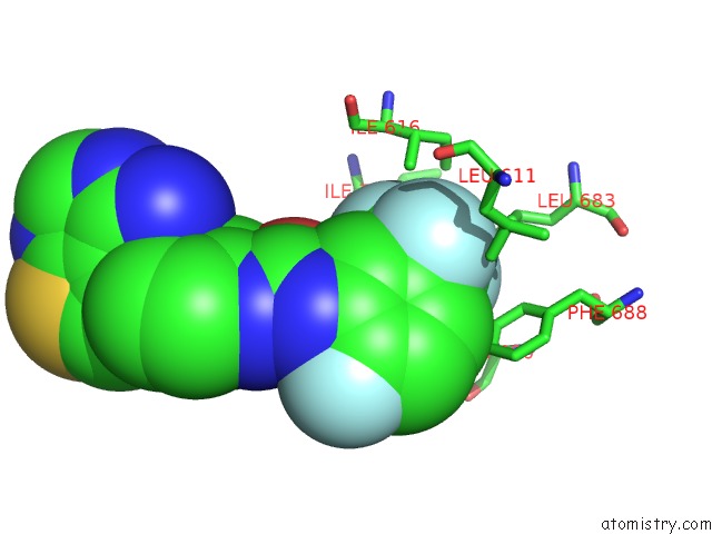
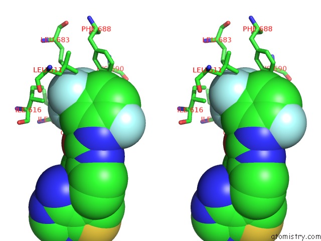
Fluorine binding site 1 out of 4 in 4at4
Go back to
Fluorine binding site 1 out
of 4 in the Crystal Structure of Trkb Kinase Domain in Complex with EX429
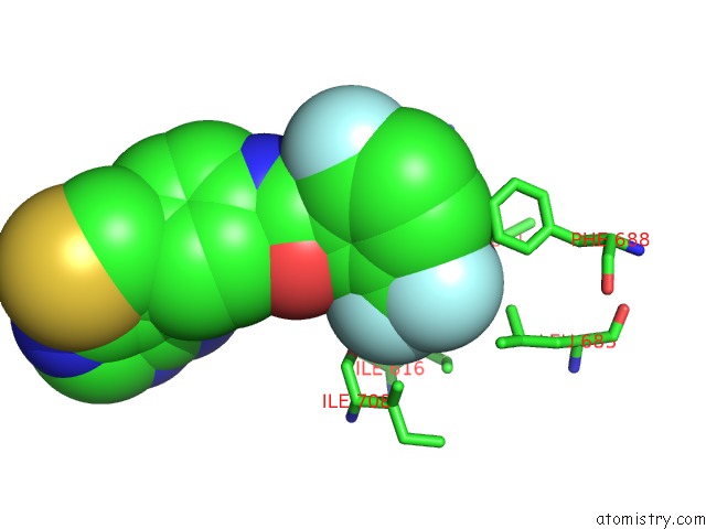
Mono view
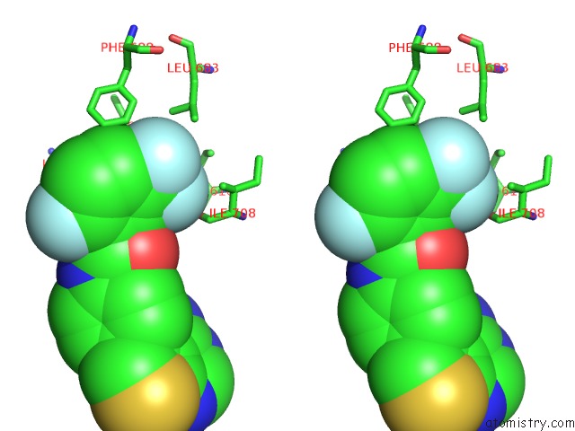
Stereo pair view
Mono view
Stereo pair view
A full contact list of Fluorine with other atoms in the F binding
site number 1 of Crystal Structure of Trkb Kinase Domain in Complex with EX429 within 5.0Å range:
|
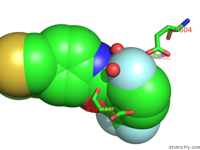
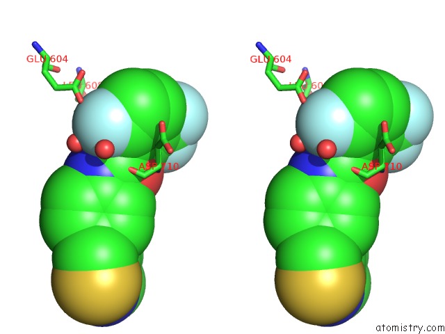
Fluorine binding site 2 out of 4 in 4at4
Go back to
Fluorine binding site 2 out
of 4 in the Crystal Structure of Trkb Kinase Domain in Complex with EX429
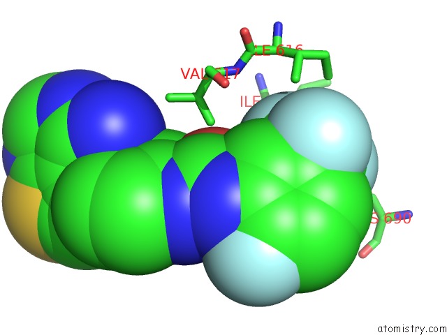
Mono view
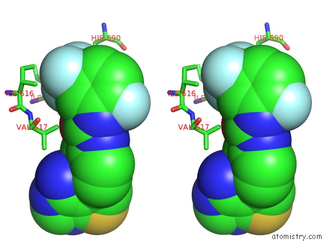
Stereo pair view
Mono view
Stereo pair view
A full contact list of Fluorine with other atoms in the F binding
site number 2 of Crystal Structure of Trkb Kinase Domain in Complex with EX429 within 5.0Å range:
|
Fluorine binding site 3 out of 4 in 4at4
Go back to
Fluorine binding site 3 out
of 4 in the Crystal Structure of Trkb Kinase Domain in Complex with EX429
Mono view
Stereo pair view
Mono view
Stereo pair view
A full contact list of Fluorine with other atoms in the F binding
site number 3 of Crystal Structure of Trkb Kinase Domain in Complex with EX429 within 5.0Å range:
|
Fluorine binding site 4 out of 4 in 4at4
Go back to
Fluorine binding site 4 out
of 4 in the Crystal Structure of Trkb Kinase Domain in Complex with EX429
Mono view
Stereo pair view
Mono view
Stereo pair view
A full contact list of Fluorine with other atoms in the F binding
site number 4 of Crystal Structure of Trkb Kinase Domain in Complex with EX429 within 5.0Å range:
|
Reference:
T.Bertrand,
M.Kothe,
J.Liu,
A.Dupuy,
A.Rak,
P.F.Berne,
S.Davis,
T.Gladysheva,
C.Valtre,
J.Y.Crenne,
M.Mathieu.
The Crystal Structures of Trka and Trkb Suggest Key Regions For Achieving Selective Inhibition. J.Mol.Biol. V. 423 439 2012.
ISSN: ISSN 0022-2836
PubMed: 22902478
DOI: 10.1016/J.JMB.2012.08.002
Page generated: Thu Aug 1 00:03:47 2024
ISSN: ISSN 0022-2836
PubMed: 22902478
DOI: 10.1016/J.JMB.2012.08.002
Last articles
Zn in 9J0NZn in 9J0O
Zn in 9J0P
Zn in 9FJX
Zn in 9EKB
Zn in 9C0F
Zn in 9CAH
Zn in 9CH0
Zn in 9CH3
Zn in 9CH1