Fluorine »
PDB 4c6l-4cqj »
4c6l »
Fluorine in PDB 4c6l: Crystal Structure of the Dihydroorotase Domain of Human Cad Bound to the Inhibitor Fluoroorotate at pH 6.0
Enzymatic activity of Crystal Structure of the Dihydroorotase Domain of Human Cad Bound to the Inhibitor Fluoroorotate at pH 6.0
All present enzymatic activity of Crystal Structure of the Dihydroorotase Domain of Human Cad Bound to the Inhibitor Fluoroorotate at pH 6.0:
3.5.2.3;
3.5.2.3;
Protein crystallography data
The structure of Crystal Structure of the Dihydroorotase Domain of Human Cad Bound to the Inhibitor Fluoroorotate at pH 6.0, PDB code: 4c6l
was solved by
S.Ramon-Maiques,
N.Lallous,
A.Grande-Garcia,
with X-Ray Crystallography technique. A brief refinement statistics is given in the table below:
| Resolution Low / High (Å) | 44.381 / 1.55 |
| Space group | C 2 2 21 |
| Cell size a, b, c (Å), α, β, γ (°) | 81.960, 158.370, 61.260, 90.00, 90.00, 90.00 |
| R / Rfree (%) | 12.37 / 15.87 |
Other elements in 4c6l:
The structure of Crystal Structure of the Dihydroorotase Domain of Human Cad Bound to the Inhibitor Fluoroorotate at pH 6.0 also contains other interesting chemical elements:
| Zinc | (Zn) | 2 atoms |
Fluorine Binding Sites:
The binding sites of Fluorine atom in the Crystal Structure of the Dihydroorotase Domain of Human Cad Bound to the Inhibitor Fluoroorotate at pH 6.0
(pdb code 4c6l). This binding sites where shown within
5.0 Angstroms radius around Fluorine atom.
In total only one binding site of Fluorine was determined in the Crystal Structure of the Dihydroorotase Domain of Human Cad Bound to the Inhibitor Fluoroorotate at pH 6.0, PDB code: 4c6l:
In total only one binding site of Fluorine was determined in the Crystal Structure of the Dihydroorotase Domain of Human Cad Bound to the Inhibitor Fluoroorotate at pH 6.0, PDB code: 4c6l:
Fluorine binding site 1 out of 1 in 4c6l
Go back to
Fluorine binding site 1 out
of 1 in the Crystal Structure of the Dihydroorotase Domain of Human Cad Bound to the Inhibitor Fluoroorotate at pH 6.0

Mono view
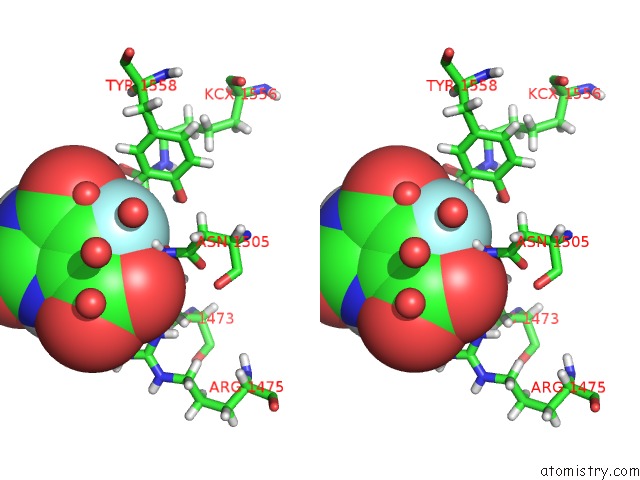
Stereo pair view
Mono view
Stereo pair view
A full contact list of Fluorine with other atoms in the F binding
site number 1 of Crystal Structure of the Dihydroorotase Domain of Human Cad Bound to the Inhibitor Fluoroorotate at pH 6.0 within 5.0Å range:
|
Reference:
A.Grande-Garcia,
N.Lallous,
C.Diaz-Tejada,
S.Ramon-Maiques.
Structure, Functional Characterization and Evolution of the Dihydroorotase Domain of Human Cad. Structure V. 22 185 2014.
ISSN: ISSN 0969-2126
PubMed: 24332717
DOI: 10.1016/J.STR.2013.10.016
Page generated: Mon Jul 14 20:55:22 2025
ISSN: ISSN 0969-2126
PubMed: 24332717
DOI: 10.1016/J.STR.2013.10.016
Last articles
F in 4RRNF in 4RPV
F in 4RM2
F in 4RLP
F in 4RK8
F in 4RLO
F in 4QZS
F in 4R88
F in 4RIO
F in 4RG0