Fluorine »
PDB 4dbu-4e28 »
4dk8 »
Fluorine in PDB 4dk8: Crystal Structure of Lxr Ligand Binding Domain in Complex with Partial Agonist 5
Enzymatic activity of Crystal Structure of Lxr Ligand Binding Domain in Complex with Partial Agonist 5
All present enzymatic activity of Crystal Structure of Lxr Ligand Binding Domain in Complex with Partial Agonist 5:
2.3.1.48;
2.3.1.48;
Protein crystallography data
The structure of Crystal Structure of Lxr Ligand Binding Domain in Complex with Partial Agonist 5, PDB code: 4dk8
was solved by
D.E.Piper,
D.J.Kopecky,
H.Xu,
with X-Ray Crystallography technique. A brief refinement statistics is given in the table below:
| Resolution Low / High (Å) | 29.73 / 2.75 |
| Space group | P 43 21 2 |
| Cell size a, b, c (Å), α, β, γ (°) | 88.145, 88.145, 198.450, 90.00, 90.00, 90.00 |
| R / Rfree (%) | 24.5 / 27.8 |
Other elements in 4dk8:
The structure of Crystal Structure of Lxr Ligand Binding Domain in Complex with Partial Agonist 5 also contains other interesting chemical elements:
| Calcium | (Ca) | 6 atoms |
Fluorine Binding Sites:
The binding sites of Fluorine atom in the Crystal Structure of Lxr Ligand Binding Domain in Complex with Partial Agonist 5
(pdb code 4dk8). This binding sites where shown within
5.0 Angstroms radius around Fluorine atom.
In total 6 binding sites of Fluorine where determined in the Crystal Structure of Lxr Ligand Binding Domain in Complex with Partial Agonist 5, PDB code: 4dk8:
Jump to Fluorine binding site number: 1; 2; 3; 4; 5; 6;
In total 6 binding sites of Fluorine where determined in the Crystal Structure of Lxr Ligand Binding Domain in Complex with Partial Agonist 5, PDB code: 4dk8:
Jump to Fluorine binding site number: 1; 2; 3; 4; 5; 6;
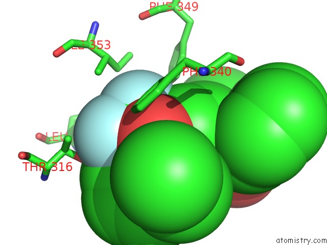
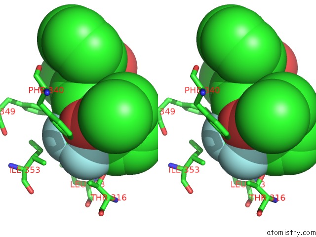
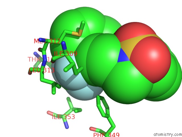
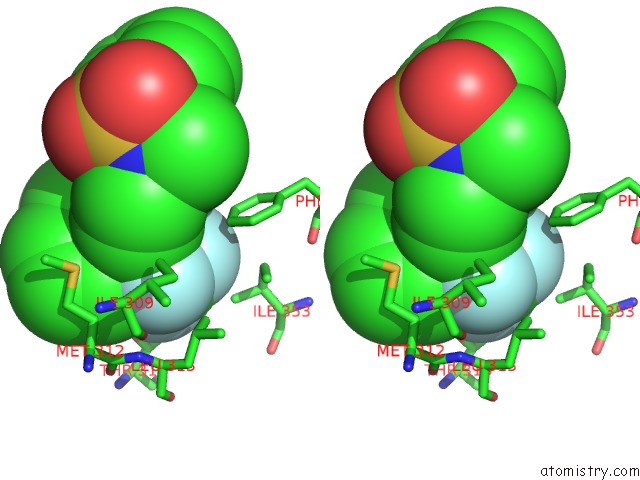
Fluorine binding site 1 out of 6 in 4dk8
Go back to
Fluorine binding site 1 out
of 6 in the Crystal Structure of Lxr Ligand Binding Domain in Complex with Partial Agonist 5
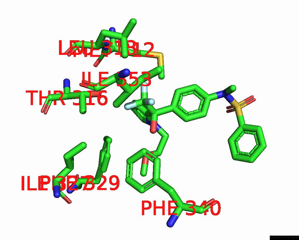
Mono view
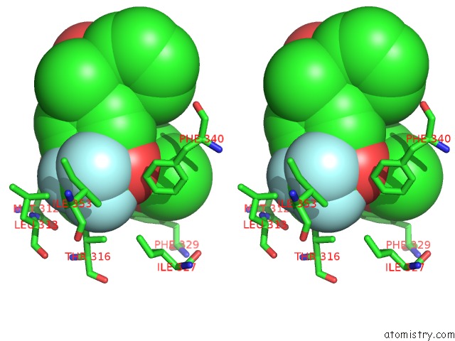
Stereo pair view
Mono view
Stereo pair view
A full contact list of Fluorine with other atoms in the F binding
site number 1 of Crystal Structure of Lxr Ligand Binding Domain in Complex with Partial Agonist 5 within 5.0Å range:
|
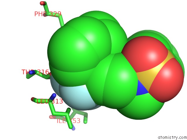
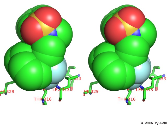
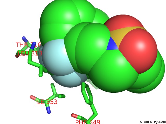
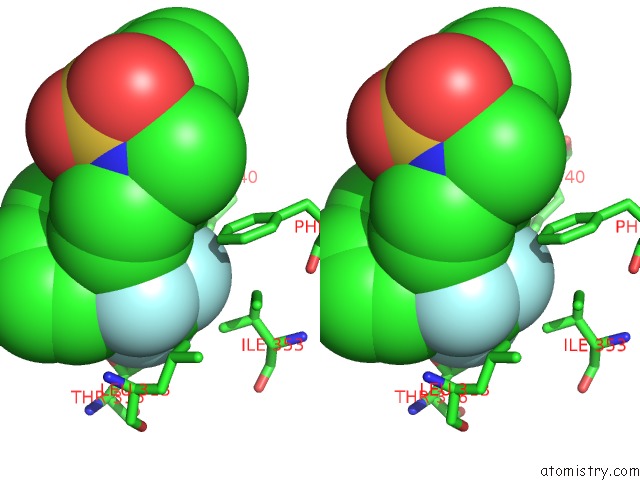
Fluorine binding site 2 out of 6 in 4dk8
Go back to
Fluorine binding site 2 out
of 6 in the Crystal Structure of Lxr Ligand Binding Domain in Complex with Partial Agonist 5
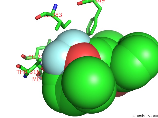
Mono view
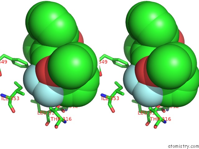
Stereo pair view
Mono view
Stereo pair view
A full contact list of Fluorine with other atoms in the F binding
site number 2 of Crystal Structure of Lxr Ligand Binding Domain in Complex with Partial Agonist 5 within 5.0Å range:
|
Fluorine binding site 3 out of 6 in 4dk8
Go back to
Fluorine binding site 3 out
of 6 in the Crystal Structure of Lxr Ligand Binding Domain in Complex with Partial Agonist 5
Mono view
Stereo pair view
Mono view
Stereo pair view
A full contact list of Fluorine with other atoms in the F binding
site number 3 of Crystal Structure of Lxr Ligand Binding Domain in Complex with Partial Agonist 5 within 5.0Å range:
|
Fluorine binding site 4 out of 6 in 4dk8
Go back to
Fluorine binding site 4 out
of 6 in the Crystal Structure of Lxr Ligand Binding Domain in Complex with Partial Agonist 5
Mono view
Stereo pair view
Mono view
Stereo pair view
A full contact list of Fluorine with other atoms in the F binding
site number 4 of Crystal Structure of Lxr Ligand Binding Domain in Complex with Partial Agonist 5 within 5.0Å range:
|
Fluorine binding site 5 out of 6 in 4dk8
Go back to
Fluorine binding site 5 out
of 6 in the Crystal Structure of Lxr Ligand Binding Domain in Complex with Partial Agonist 5
Mono view
Stereo pair view
Mono view
Stereo pair view
A full contact list of Fluorine with other atoms in the F binding
site number 5 of Crystal Structure of Lxr Ligand Binding Domain in Complex with Partial Agonist 5 within 5.0Å range:
|
Fluorine binding site 6 out of 6 in 4dk8
Go back to
Fluorine binding site 6 out
of 6 in the Crystal Structure of Lxr Ligand Binding Domain in Complex with Partial Agonist 5
Mono view
Stereo pair view
Mono view
Stereo pair view
A full contact list of Fluorine with other atoms in the F binding
site number 6 of Crystal Structure of Lxr Ligand Binding Domain in Complex with Partial Agonist 5 within 5.0Å range:
|
Reference:
D.J.Kopecky,
X.Y.Jiao,
B.Fisher,
S.Mckendry,
M.Labelle,
D.E.Piper,
P.Coward,
A.K.Shiau,
P.Escaron,
J.Danao,
A.Chai,
J.Jaen,
F.Kayser.
Discovery of A New Binding Mode For A Series of Liver X Receptor Agonists. Bioorg.Med.Chem.Lett. V. 22 2407 2012.
ISSN: ISSN 0960-894X
PubMed: 22406115
DOI: 10.1016/J.BMCL.2012.02.028
Page generated: Mon Jul 14 21:11:44 2025
ISSN: ISSN 0960-894X
PubMed: 22406115
DOI: 10.1016/J.BMCL.2012.02.028
Last articles
F in 7KYVF in 7KYT
F in 7KYB
F in 7KYC
F in 7KYA
F in 7KY5
F in 7KXW
F in 7KY9
F in 7KWA
F in 7KXT