Fluorine »
PDB 5sb4-5sjr »
5sif »
Fluorine in PDB 5sif: Crystal Structure of Human Phosphodiesterase 10 in Complex with 3-[2- (2,5-Difluorophenyl)Pyrazol-3-Yl]-1-(3-Methylsulfonylphenyl) Pyridazin-4-One
Enzymatic activity of Crystal Structure of Human Phosphodiesterase 10 in Complex with 3-[2- (2,5-Difluorophenyl)Pyrazol-3-Yl]-1-(3-Methylsulfonylphenyl) Pyridazin-4-One
All present enzymatic activity of Crystal Structure of Human Phosphodiesterase 10 in Complex with 3-[2- (2,5-Difluorophenyl)Pyrazol-3-Yl]-1-(3-Methylsulfonylphenyl) Pyridazin-4-One:
3.1.4.17;
3.1.4.17;
Protein crystallography data
The structure of Crystal Structure of Human Phosphodiesterase 10 in Complex with 3-[2- (2,5-Difluorophenyl)Pyrazol-3-Yl]-1-(3-Methylsulfonylphenyl) Pyridazin-4-One, PDB code: 5sif
was solved by
C.Joseph,
J.Benz,
A.Flohr,
M.Rogers-Evans,
M.G.Rudolph,
with X-Ray Crystallography technique. A brief refinement statistics is given in the table below:
| Resolution Low / High (Å) | 41.45 / 2.20 |
| Space group | H 3 |
| Cell size a, b, c (Å), α, β, γ (°) | 135.135, 135.135, 235.085, 90, 90, 120 |
| R / Rfree (%) | 18.2 / 23.1 |
Other elements in 5sif:
The structure of Crystal Structure of Human Phosphodiesterase 10 in Complex with 3-[2- (2,5-Difluorophenyl)Pyrazol-3-Yl]-1-(3-Methylsulfonylphenyl) Pyridazin-4-One also contains other interesting chemical elements:
| Zinc | (Zn) | 4 atoms |
| Chlorine | (Cl) | 1 atom |
| Praseodymium | (Pr) | 8 atoms |
| Magnesium | (Mg) | 4 atoms |
Fluorine Binding Sites:
The binding sites of Fluorine atom in the Crystal Structure of Human Phosphodiesterase 10 in Complex with 3-[2- (2,5-Difluorophenyl)Pyrazol-3-Yl]-1-(3-Methylsulfonylphenyl) Pyridazin-4-One
(pdb code 5sif). This binding sites where shown within
5.0 Angstroms radius around Fluorine atom.
In total 8 binding sites of Fluorine where determined in the Crystal Structure of Human Phosphodiesterase 10 in Complex with 3-[2- (2,5-Difluorophenyl)Pyrazol-3-Yl]-1-(3-Methylsulfonylphenyl) Pyridazin-4-One, PDB code: 5sif:
Jump to Fluorine binding site number: 1; 2; 3; 4; 5; 6; 7; 8;
In total 8 binding sites of Fluorine where determined in the Crystal Structure of Human Phosphodiesterase 10 in Complex with 3-[2- (2,5-Difluorophenyl)Pyrazol-3-Yl]-1-(3-Methylsulfonylphenyl) Pyridazin-4-One, PDB code: 5sif:
Jump to Fluorine binding site number: 1; 2; 3; 4; 5; 6; 7; 8;
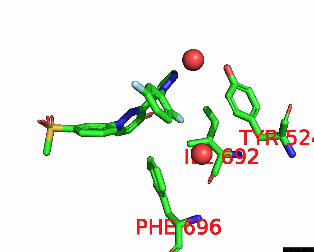
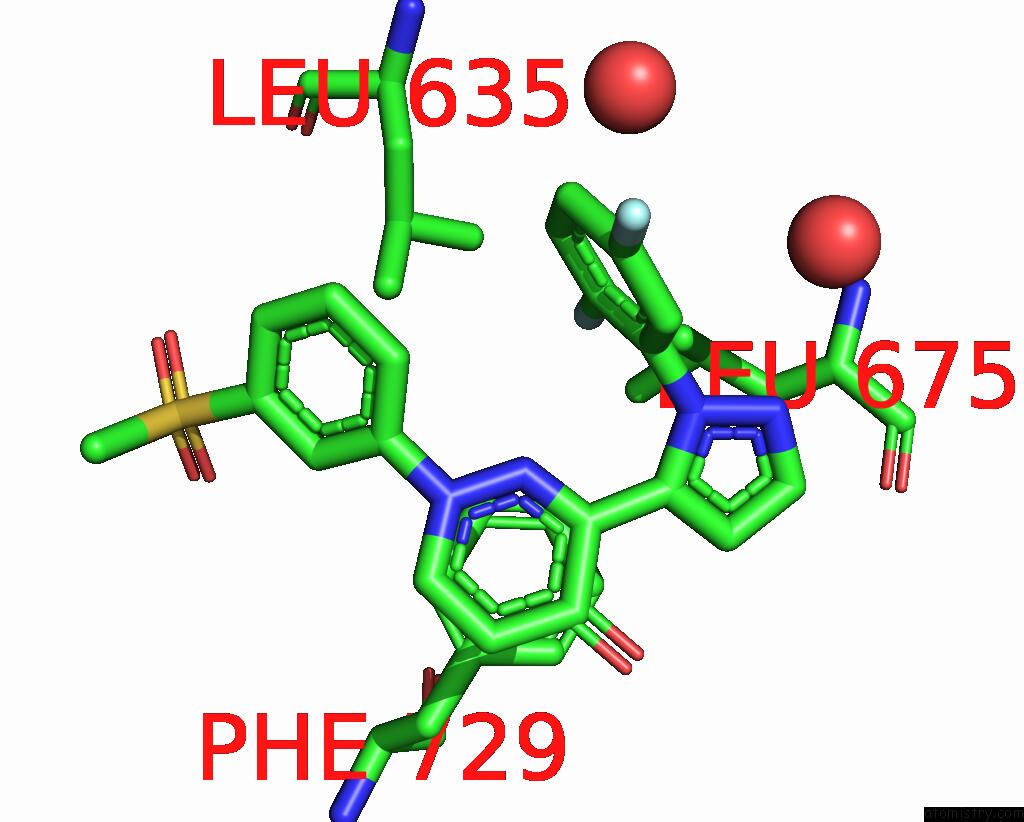
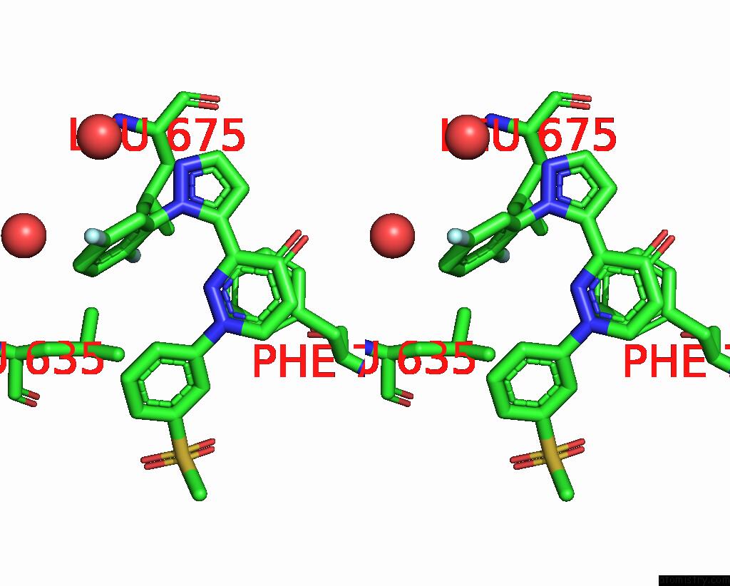
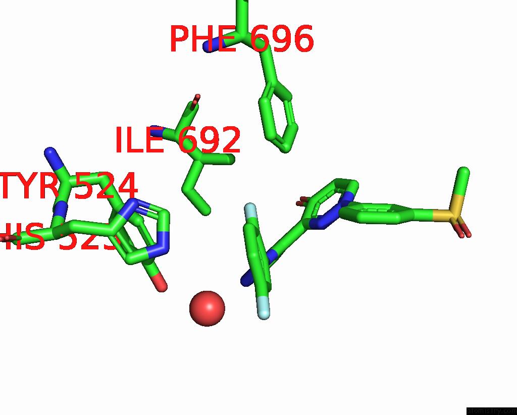
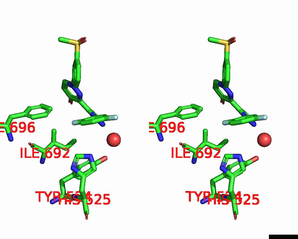
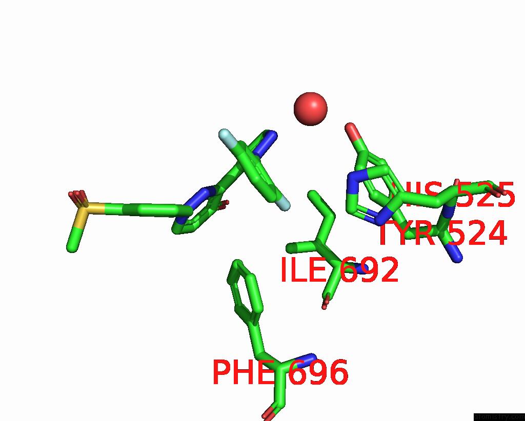
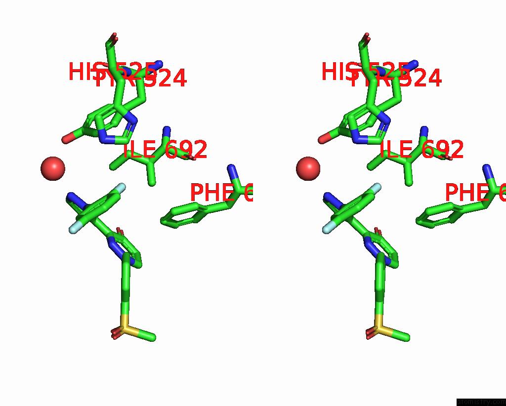
Fluorine binding site 1 out of 8 in 5sif
Go back to
Fluorine binding site 1 out
of 8 in the Crystal Structure of Human Phosphodiesterase 10 in Complex with 3-[2- (2,5-Difluorophenyl)Pyrazol-3-Yl]-1-(3-Methylsulfonylphenyl) Pyridazin-4-One
Mono view
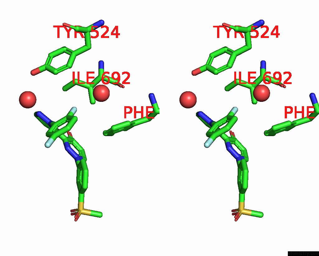
Stereo pair view
Mono view
Stereo pair view
A full contact list of Fluorine with other atoms in the F binding
site number 1 of Crystal Structure of Human Phosphodiesterase 10 in Complex with 3-[2- (2,5-Difluorophenyl)Pyrazol-3-Yl]-1-(3-Methylsulfonylphenyl) Pyridazin-4-One within 5.0Å range:
|
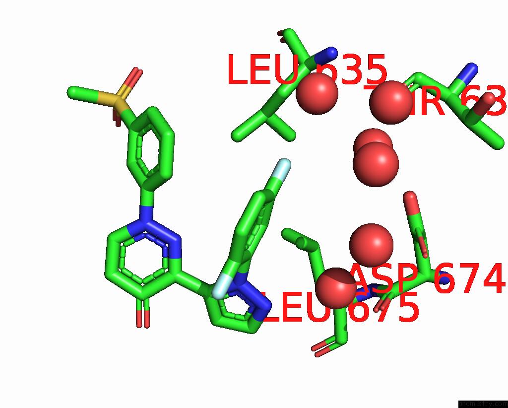
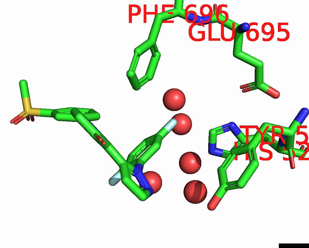
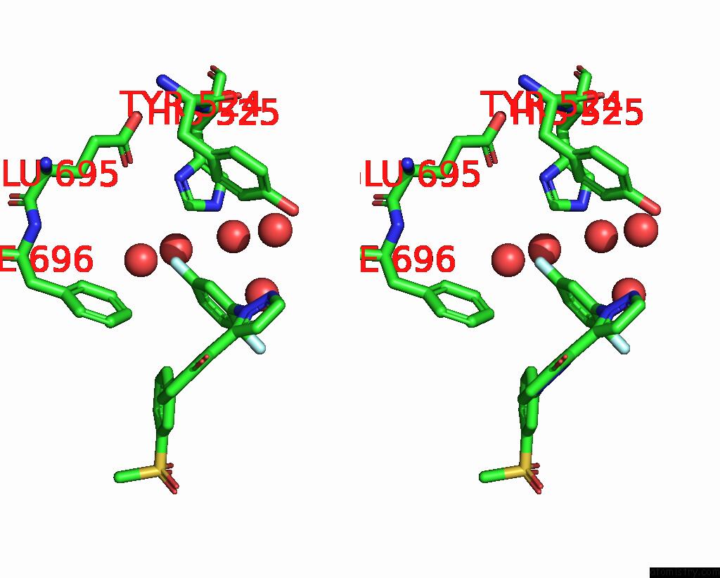
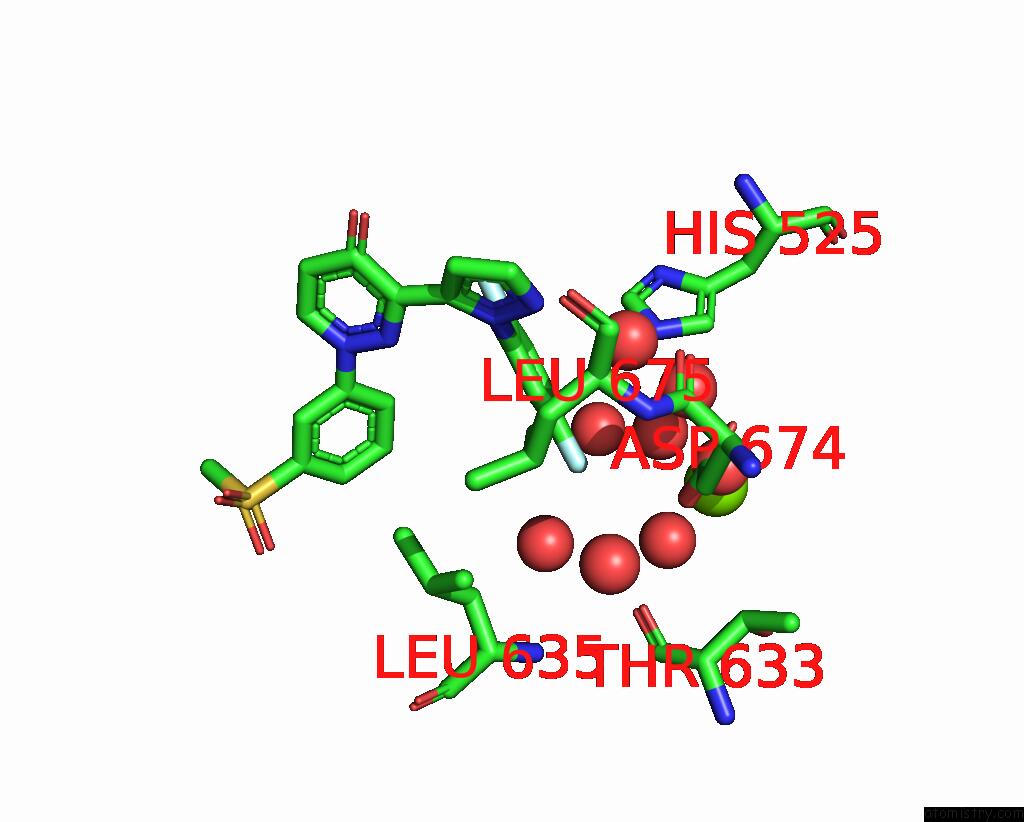
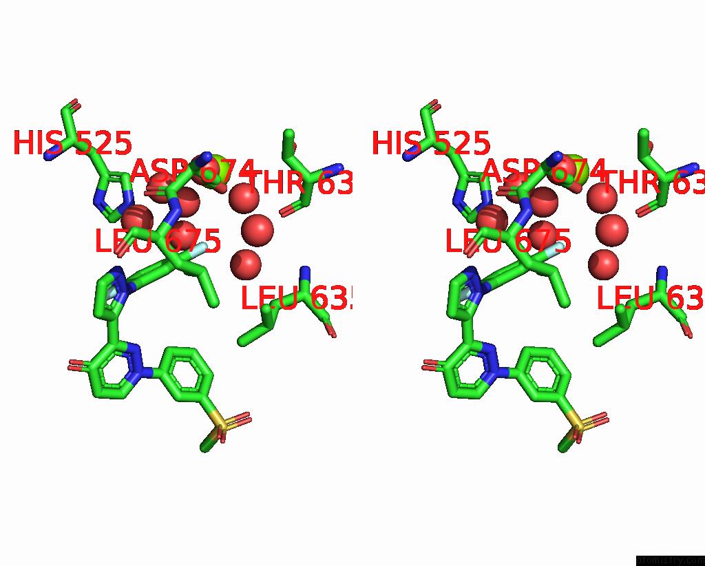
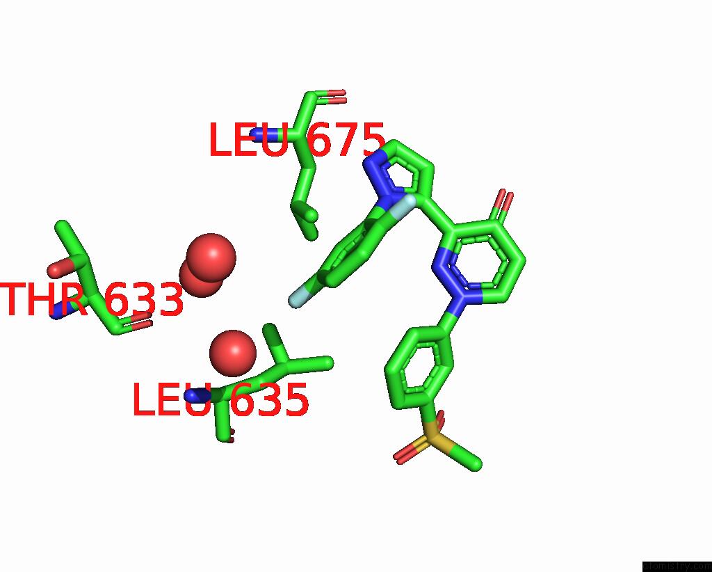
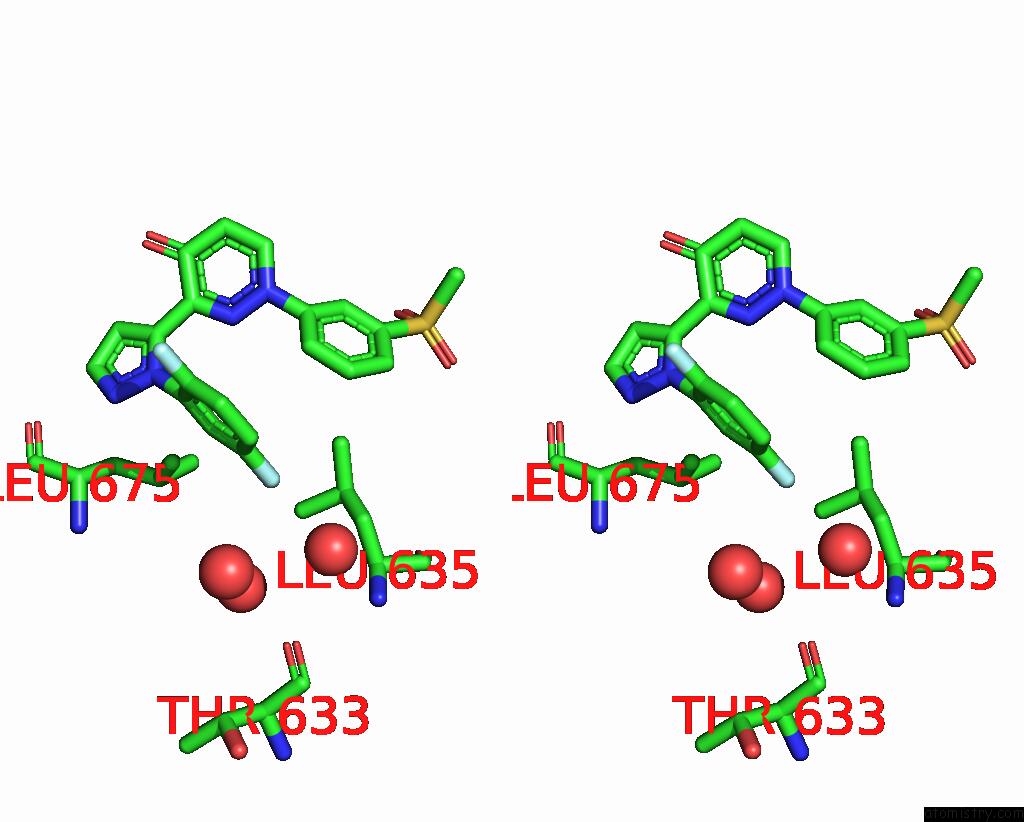
Fluorine binding site 2 out of 8 in 5sif
Go back to
Fluorine binding site 2 out
of 8 in the Crystal Structure of Human Phosphodiesterase 10 in Complex with 3-[2- (2,5-Difluorophenyl)Pyrazol-3-Yl]-1-(3-Methylsulfonylphenyl) Pyridazin-4-One
Mono view
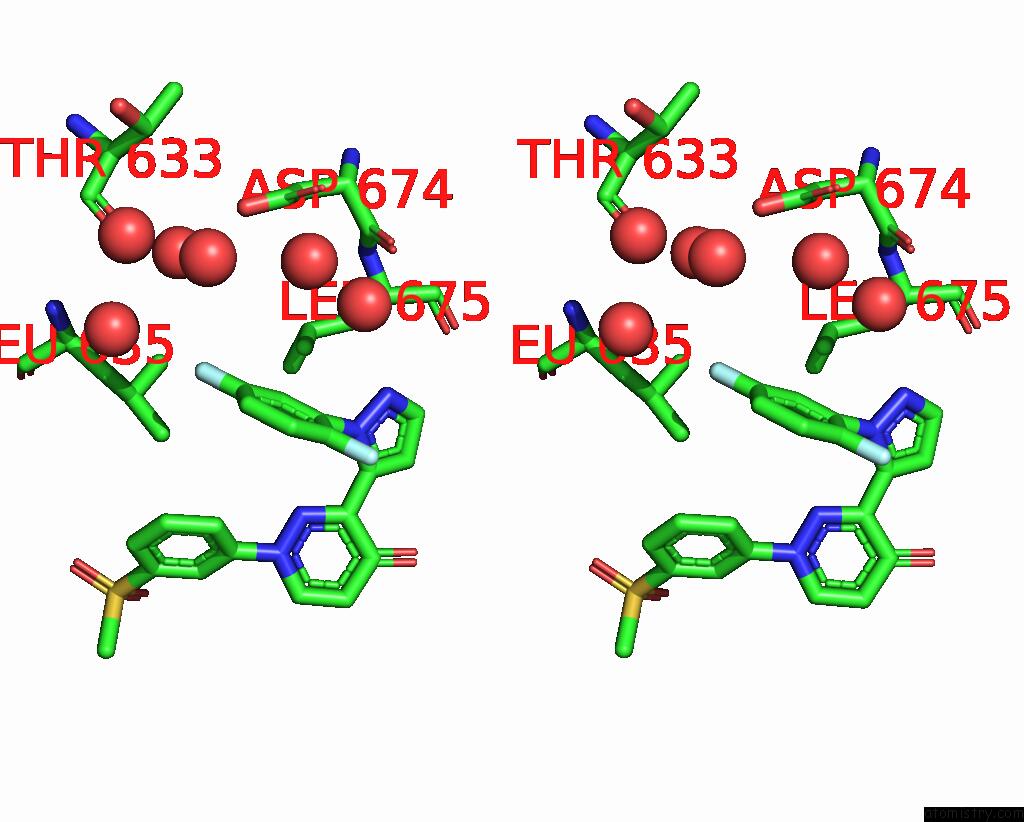
Stereo pair view
Mono view
Stereo pair view
A full contact list of Fluorine with other atoms in the F binding
site number 2 of Crystal Structure of Human Phosphodiesterase 10 in Complex with 3-[2- (2,5-Difluorophenyl)Pyrazol-3-Yl]-1-(3-Methylsulfonylphenyl) Pyridazin-4-One within 5.0Å range:
|
Fluorine binding site 3 out of 8 in 5sif
Go back to
Fluorine binding site 3 out
of 8 in the Crystal Structure of Human Phosphodiesterase 10 in Complex with 3-[2- (2,5-Difluorophenyl)Pyrazol-3-Yl]-1-(3-Methylsulfonylphenyl) Pyridazin-4-One
Mono view
Stereo pair view
Mono view
Stereo pair view
A full contact list of Fluorine with other atoms in the F binding
site number 3 of Crystal Structure of Human Phosphodiesterase 10 in Complex with 3-[2- (2,5-Difluorophenyl)Pyrazol-3-Yl]-1-(3-Methylsulfonylphenyl) Pyridazin-4-One within 5.0Å range:
|
Fluorine binding site 4 out of 8 in 5sif
Go back to
Fluorine binding site 4 out
of 8 in the Crystal Structure of Human Phosphodiesterase 10 in Complex with 3-[2- (2,5-Difluorophenyl)Pyrazol-3-Yl]-1-(3-Methylsulfonylphenyl) Pyridazin-4-One
Mono view
Stereo pair view
Mono view
Stereo pair view
A full contact list of Fluorine with other atoms in the F binding
site number 4 of Crystal Structure of Human Phosphodiesterase 10 in Complex with 3-[2- (2,5-Difluorophenyl)Pyrazol-3-Yl]-1-(3-Methylsulfonylphenyl) Pyridazin-4-One within 5.0Å range:
|
Fluorine binding site 5 out of 8 in 5sif
Go back to
Fluorine binding site 5 out
of 8 in the Crystal Structure of Human Phosphodiesterase 10 in Complex with 3-[2- (2,5-Difluorophenyl)Pyrazol-3-Yl]-1-(3-Methylsulfonylphenyl) Pyridazin-4-One
Mono view
Stereo pair view
Mono view
Stereo pair view
A full contact list of Fluorine with other atoms in the F binding
site number 5 of Crystal Structure of Human Phosphodiesterase 10 in Complex with 3-[2- (2,5-Difluorophenyl)Pyrazol-3-Yl]-1-(3-Methylsulfonylphenyl) Pyridazin-4-One within 5.0Å range:
|
Fluorine binding site 6 out of 8 in 5sif
Go back to
Fluorine binding site 6 out
of 8 in the Crystal Structure of Human Phosphodiesterase 10 in Complex with 3-[2- (2,5-Difluorophenyl)Pyrazol-3-Yl]-1-(3-Methylsulfonylphenyl) Pyridazin-4-One
Mono view
Stereo pair view
Mono view
Stereo pair view
A full contact list of Fluorine with other atoms in the F binding
site number 6 of Crystal Structure of Human Phosphodiesterase 10 in Complex with 3-[2- (2,5-Difluorophenyl)Pyrazol-3-Yl]-1-(3-Methylsulfonylphenyl) Pyridazin-4-One within 5.0Å range:
|
Fluorine binding site 7 out of 8 in 5sif
Go back to
Fluorine binding site 7 out
of 8 in the Crystal Structure of Human Phosphodiesterase 10 in Complex with 3-[2- (2,5-Difluorophenyl)Pyrazol-3-Yl]-1-(3-Methylsulfonylphenyl) Pyridazin-4-One
Mono view
Stereo pair view
Mono view
Stereo pair view
A full contact list of Fluorine with other atoms in the F binding
site number 7 of Crystal Structure of Human Phosphodiesterase 10 in Complex with 3-[2- (2,5-Difluorophenyl)Pyrazol-3-Yl]-1-(3-Methylsulfonylphenyl) Pyridazin-4-One within 5.0Å range:
|
Fluorine binding site 8 out of 8 in 5sif
Go back to
Fluorine binding site 8 out
of 8 in the Crystal Structure of Human Phosphodiesterase 10 in Complex with 3-[2- (2,5-Difluorophenyl)Pyrazol-3-Yl]-1-(3-Methylsulfonylphenyl) Pyridazin-4-One
Mono view
Stereo pair view
Mono view
Stereo pair view
A full contact list of Fluorine with other atoms in the F binding
site number 8 of Crystal Structure of Human Phosphodiesterase 10 in Complex with 3-[2- (2,5-Difluorophenyl)Pyrazol-3-Yl]-1-(3-Methylsulfonylphenyl) Pyridazin-4-One within 5.0Å range:
|
Reference:
A.Flohr,
D.Schlatter,
B.Kuhn,
M.G.Rudolph.
Crystal Structure of A Human Phosphodiesterase 10 Complex To Be Published.
Page generated: Tue Jul 15 07:17:53 2025
Last articles
F in 6NG7F in 6NG6
F in 6NG5
F in 6NG4
F in 6NG1
F in 6NG2
F in 6NFZ
F in 6NG0
F in 6NDY
F in 6NFY