Fluorine »
PDB 5sk2-5soj »
5so3 »
Fluorine in PDB 5so3: Pandda Analysis Group Deposition -- Crystal Structure of Pseudomonas Aeruginosa Fabf-C164Q Mutant Protein in Complex with JKH100B
Enzymatic activity of Pandda Analysis Group Deposition -- Crystal Structure of Pseudomonas Aeruginosa Fabf-C164Q Mutant Protein in Complex with JKH100B
All present enzymatic activity of Pandda Analysis Group Deposition -- Crystal Structure of Pseudomonas Aeruginosa Fabf-C164Q Mutant Protein in Complex with JKH100B:
2.3.1.179;
2.3.1.179;
Protein crystallography data
The structure of Pandda Analysis Group Deposition -- Crystal Structure of Pseudomonas Aeruginosa Fabf-C164Q Mutant Protein in Complex with JKH100B, PDB code: 5so3
was solved by
R.Brenk,
C.Georgiou,
with X-Ray Crystallography technique. A brief refinement statistics is given in the table below:
| Resolution Low / High (Å) | 49.08 / 1.49 |
| Space group | C 1 2 1 |
| Cell size a, b, c (Å), α, β, γ (°) | 137.77, 65.44, 84.51, 90, 93.68, 90 |
| R / Rfree (%) | 17 / 19.8 |
Fluorine Binding Sites:
The binding sites of Fluorine atom in the Pandda Analysis Group Deposition -- Crystal Structure of Pseudomonas Aeruginosa Fabf-C164Q Mutant Protein in Complex with JKH100B
(pdb code 5so3). This binding sites where shown within
5.0 Angstroms radius around Fluorine atom.
In total 2 binding sites of Fluorine where determined in the Pandda Analysis Group Deposition -- Crystal Structure of Pseudomonas Aeruginosa Fabf-C164Q Mutant Protein in Complex with JKH100B, PDB code: 5so3:
Jump to Fluorine binding site number: 1; 2;
In total 2 binding sites of Fluorine where determined in the Pandda Analysis Group Deposition -- Crystal Structure of Pseudomonas Aeruginosa Fabf-C164Q Mutant Protein in Complex with JKH100B, PDB code: 5so3:
Jump to Fluorine binding site number: 1; 2;
Fluorine binding site 1 out of 2 in 5so3
Go back to
Fluorine binding site 1 out
of 2 in the Pandda Analysis Group Deposition -- Crystal Structure of Pseudomonas Aeruginosa Fabf-C164Q Mutant Protein in Complex with JKH100B
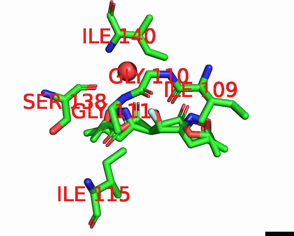
Mono view
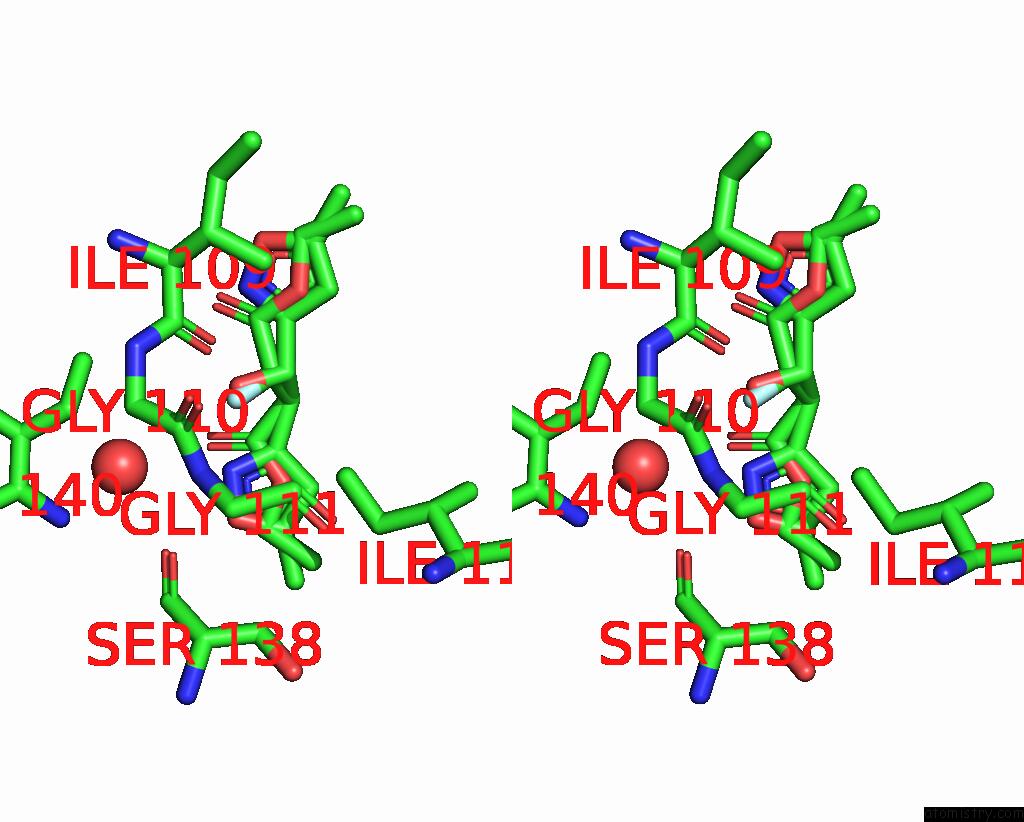
Stereo pair view
Mono view
Stereo pair view
A full contact list of Fluorine with other atoms in the F binding
site number 1 of Pandda Analysis Group Deposition -- Crystal Structure of Pseudomonas Aeruginosa Fabf-C164Q Mutant Protein in Complex with JKH100B within 5.0Å range:
|
Fluorine binding site 2 out of 2 in 5so3
Go back to
Fluorine binding site 2 out
of 2 in the Pandda Analysis Group Deposition -- Crystal Structure of Pseudomonas Aeruginosa Fabf-C164Q Mutant Protein in Complex with JKH100B
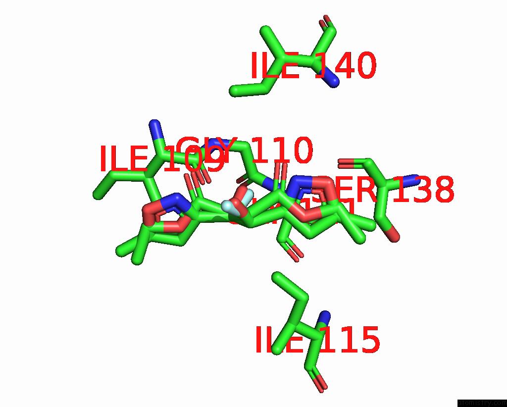
Mono view
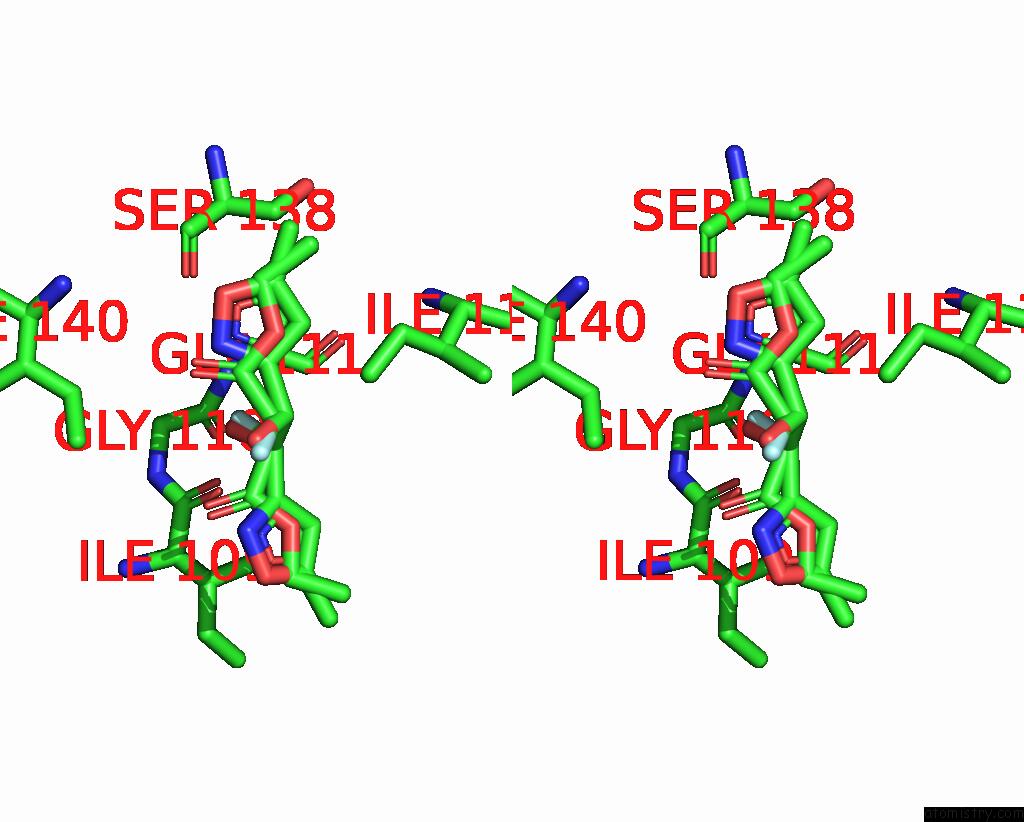
Stereo pair view
Mono view
Stereo pair view
A full contact list of Fluorine with other atoms in the F binding
site number 2 of Pandda Analysis Group Deposition -- Crystal Structure of Pseudomonas Aeruginosa Fabf-C164Q Mutant Protein in Complex with JKH100B within 5.0Å range:
|
Reference:
R.Brenk,
C.Georgiou.
Pandda Analysis Group Deposition To Be Published.
Page generated: Tue Jul 15 07:31:55 2025
Last articles
F in 6I3SF in 6I03
F in 6I15
F in 6I0L
F in 6HWV
F in 6HU3
F in 6HS2
F in 6HWU
F in 6HPU
F in 6HWT