Fluorine »
PDB 6b3e-6bkw »
6b7e »
Fluorine in PDB 6b7e: Crystal Structure of E.Coli Phosphopantetheine Adenylyltransferase (Ppat/Coad) in Complex with (R)-4-(5-(Difluoromethyl)-1H-Imidazol-1- Yl)-3,3-Dimethylisochroman-1-One
Enzymatic activity of Crystal Structure of E.Coli Phosphopantetheine Adenylyltransferase (Ppat/Coad) in Complex with (R)-4-(5-(Difluoromethyl)-1H-Imidazol-1- Yl)-3,3-Dimethylisochroman-1-One
All present enzymatic activity of Crystal Structure of E.Coli Phosphopantetheine Adenylyltransferase (Ppat/Coad) in Complex with (R)-4-(5-(Difluoromethyl)-1H-Imidazol-1- Yl)-3,3-Dimethylisochroman-1-One:
2.7.7.3;
2.7.7.3;
Protein crystallography data
The structure of Crystal Structure of E.Coli Phosphopantetheine Adenylyltransferase (Ppat/Coad) in Complex with (R)-4-(5-(Difluoromethyl)-1H-Imidazol-1- Yl)-3,3-Dimethylisochroman-1-One, PDB code: 6b7e
was solved by
A.W.Proudfoot,
D.Bussiere,
A.Lingel,
with X-Ray Crystallography technique. A brief refinement statistics is given in the table below:
| Resolution Low / High (Å) | 30.29 / 2.10 |
| Space group | I 2 3 |
| Cell size a, b, c (Å), α, β, γ (°) | 135.480, 135.480, 135.480, 90.00, 90.00, 90.00 |
| R / Rfree (%) | 17.1 / 20.6 |
Fluorine Binding Sites:
The binding sites of Fluorine atom in the Crystal Structure of E.Coli Phosphopantetheine Adenylyltransferase (Ppat/Coad) in Complex with (R)-4-(5-(Difluoromethyl)-1H-Imidazol-1- Yl)-3,3-Dimethylisochroman-1-One
(pdb code 6b7e). This binding sites where shown within
5.0 Angstroms radius around Fluorine atom.
In total 2 binding sites of Fluorine where determined in the Crystal Structure of E.Coli Phosphopantetheine Adenylyltransferase (Ppat/Coad) in Complex with (R)-4-(5-(Difluoromethyl)-1H-Imidazol-1- Yl)-3,3-Dimethylisochroman-1-One, PDB code: 6b7e:
Jump to Fluorine binding site number: 1; 2;
In total 2 binding sites of Fluorine where determined in the Crystal Structure of E.Coli Phosphopantetheine Adenylyltransferase (Ppat/Coad) in Complex with (R)-4-(5-(Difluoromethyl)-1H-Imidazol-1- Yl)-3,3-Dimethylisochroman-1-One, PDB code: 6b7e:
Jump to Fluorine binding site number: 1; 2;
Fluorine binding site 1 out of 2 in 6b7e
Go back to
Fluorine binding site 1 out
of 2 in the Crystal Structure of E.Coli Phosphopantetheine Adenylyltransferase (Ppat/Coad) in Complex with (R)-4-(5-(Difluoromethyl)-1H-Imidazol-1- Yl)-3,3-Dimethylisochroman-1-One
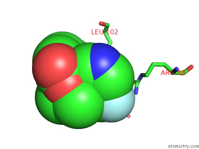
Mono view
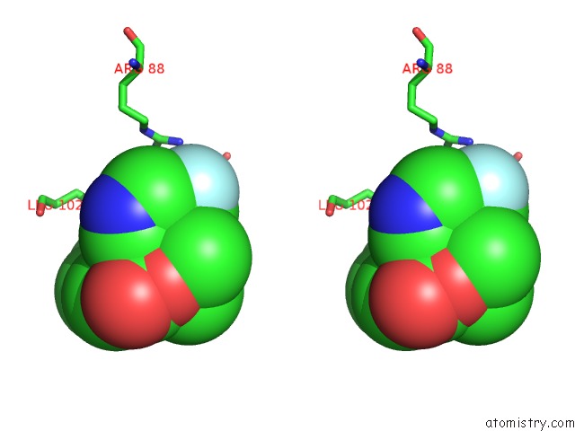
Stereo pair view
Mono view
Stereo pair view
A full contact list of Fluorine with other atoms in the F binding
site number 1 of Crystal Structure of E.Coli Phosphopantetheine Adenylyltransferase (Ppat/Coad) in Complex with (R)-4-(5-(Difluoromethyl)-1H-Imidazol-1- Yl)-3,3-Dimethylisochroman-1-One within 5.0Å range:
|
Fluorine binding site 2 out of 2 in 6b7e
Go back to
Fluorine binding site 2 out
of 2 in the Crystal Structure of E.Coli Phosphopantetheine Adenylyltransferase (Ppat/Coad) in Complex with (R)-4-(5-(Difluoromethyl)-1H-Imidazol-1- Yl)-3,3-Dimethylisochroman-1-One
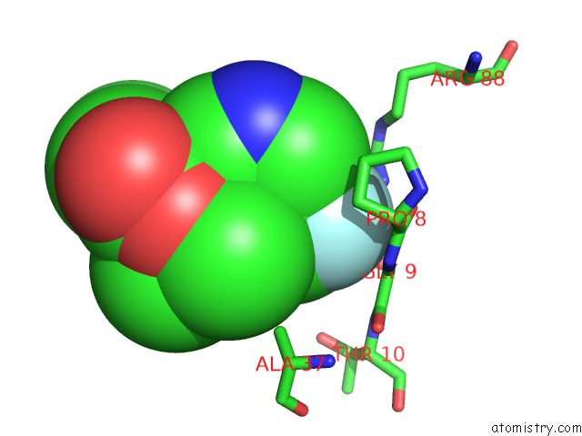
Mono view
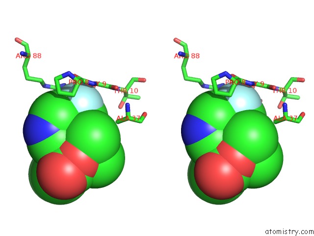
Stereo pair view
Mono view
Stereo pair view
A full contact list of Fluorine with other atoms in the F binding
site number 2 of Crystal Structure of E.Coli Phosphopantetheine Adenylyltransferase (Ppat/Coad) in Complex with (R)-4-(5-(Difluoromethyl)-1H-Imidazol-1- Yl)-3,3-Dimethylisochroman-1-One within 5.0Å range:
|
Reference:
A.Proudfoot,
D.E.Bussiere,
A.Lingel.
High-Confidence Protein-Ligand Complex Modeling By uc(Nmr)-Guided Docking Enables Early Hit Optimization. J. Am. Chem. Soc. V. 139 17824 2017.
ISSN: ESSN 1520-5126
PubMed: 29190085
DOI: 10.1021/JACS.7B07171
Page generated: Tue Jul 15 09:58:12 2025
ISSN: ESSN 1520-5126
PubMed: 29190085
DOI: 10.1021/JACS.7B07171
Last articles
F in 7JHWF in 7JHD
F in 7I18
F in 7I2F
F in 7I2M
F in 7I2A
F in 7I2D
F in 7HNS
F in 7HOG
F in 7HO4