Fluorine »
PDB 6bkz-6c0t »
6c0t »
Fluorine in PDB 6c0t: Crystal Structure of Cgmp-Dependent Protein Kinase Ialpha (Pkg Ialpha) Catalytic Domain Bound with N46
Enzymatic activity of Crystal Structure of Cgmp-Dependent Protein Kinase Ialpha (Pkg Ialpha) Catalytic Domain Bound with N46
All present enzymatic activity of Crystal Structure of Cgmp-Dependent Protein Kinase Ialpha (Pkg Ialpha) Catalytic Domain Bound with N46:
2.7.11.12;
2.7.11.12;
Protein crystallography data
The structure of Crystal Structure of Cgmp-Dependent Protein Kinase Ialpha (Pkg Ialpha) Catalytic Domain Bound with N46, PDB code: 6c0t
was solved by
L.Qin,
B.Sankaran,
C.Kim,
with X-Ray Crystallography technique. A brief refinement statistics is given in the table below:
| Resolution Low / High (Å) | 42.87 / 1.98 |
| Space group | P 42 |
| Cell size a, b, c (Å), α, β, γ (°) | 85.730, 85.730, 50.664, 90.00, 90.00, 90.00 |
| R / Rfree (%) | 20.7 / 24.4 |
Fluorine Binding Sites:
The binding sites of Fluorine atom in the Crystal Structure of Cgmp-Dependent Protein Kinase Ialpha (Pkg Ialpha) Catalytic Domain Bound with N46
(pdb code 6c0t). This binding sites where shown within
5.0 Angstroms radius around Fluorine atom.
In total only one binding site of Fluorine was determined in the Crystal Structure of Cgmp-Dependent Protein Kinase Ialpha (Pkg Ialpha) Catalytic Domain Bound with N46, PDB code: 6c0t:
In total only one binding site of Fluorine was determined in the Crystal Structure of Cgmp-Dependent Protein Kinase Ialpha (Pkg Ialpha) Catalytic Domain Bound with N46, PDB code: 6c0t:
Fluorine binding site 1 out of 1 in 6c0t
Go back to
Fluorine binding site 1 out
of 1 in the Crystal Structure of Cgmp-Dependent Protein Kinase Ialpha (Pkg Ialpha) Catalytic Domain Bound with N46
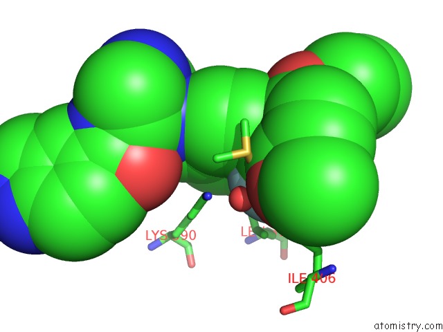
Mono view
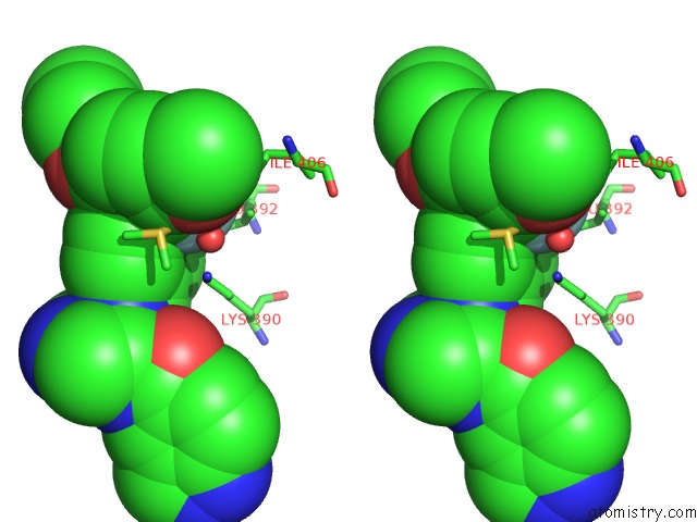
Stereo pair view
Mono view
Stereo pair view
A full contact list of Fluorine with other atoms in the F binding
site number 1 of Crystal Structure of Cgmp-Dependent Protein Kinase Ialpha (Pkg Ialpha) Catalytic Domain Bound with N46 within 5.0Å range:
|
Reference:
L.Qin,
B.Sankaran,
S.Aminzai,
D.E.Casteel,
C.Kim.
Structural Basis For Selective Inhibition of Human Pkg I Alpha By the Balanol-Like Compound N46. J. Biol. Chem. V. 293 10985 2018.
ISSN: ESSN 1083-351X
PubMed: 29769318
DOI: 10.1074/JBC.RA118.002427
Page generated: Tue Jul 15 10:18:56 2025
ISSN: ESSN 1083-351X
PubMed: 29769318
DOI: 10.1074/JBC.RA118.002427
Last articles
F in 6UGRF in 6UD8
F in 6UFX
F in 6UD4
F in 6UEL
F in 6UDZ
F in 6UDM
F in 6UCX
F in 6UCB
F in 6UD2