Fluorine »
PDB 6c0u-6cr2 »
6cqf »
Fluorine in PDB 6cqf: Crystal Structure of HPK1 in Complex An Inhibitor G1858
Enzymatic activity of Crystal Structure of HPK1 in Complex An Inhibitor G1858
All present enzymatic activity of Crystal Structure of HPK1 in Complex An Inhibitor G1858:
2.7.11.1;
2.7.11.1;
Protein crystallography data
The structure of Crystal Structure of HPK1 in Complex An Inhibitor G1858, PDB code: 6cqf
was solved by
P.Wu,
I.Lehoux,
K.Mortara,
Y.Franke,
B.K.Chan,
W.Wang,
with X-Ray Crystallography technique. A brief refinement statistics is given in the table below:
| Resolution Low / High (Å) | 33.64 / 2.25 |
| Space group | C 2 2 21 |
| Cell size a, b, c (Å), α, β, γ (°) | 91.868, 98.803, 77.100, 90.00, 90.00, 90.00 |
| R / Rfree (%) | 20.8 / 25.5 |
Fluorine Binding Sites:
The binding sites of Fluorine atom in the Crystal Structure of HPK1 in Complex An Inhibitor G1858
(pdb code 6cqf). This binding sites where shown within
5.0 Angstroms radius around Fluorine atom.
In total 2 binding sites of Fluorine where determined in the Crystal Structure of HPK1 in Complex An Inhibitor G1858, PDB code: 6cqf:
Jump to Fluorine binding site number: 1; 2;
In total 2 binding sites of Fluorine where determined in the Crystal Structure of HPK1 in Complex An Inhibitor G1858, PDB code: 6cqf:
Jump to Fluorine binding site number: 1; 2;
Fluorine binding site 1 out of 2 in 6cqf
Go back to
Fluorine binding site 1 out
of 2 in the Crystal Structure of HPK1 in Complex An Inhibitor G1858
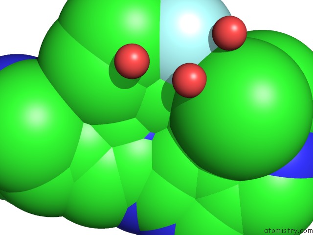
Mono view
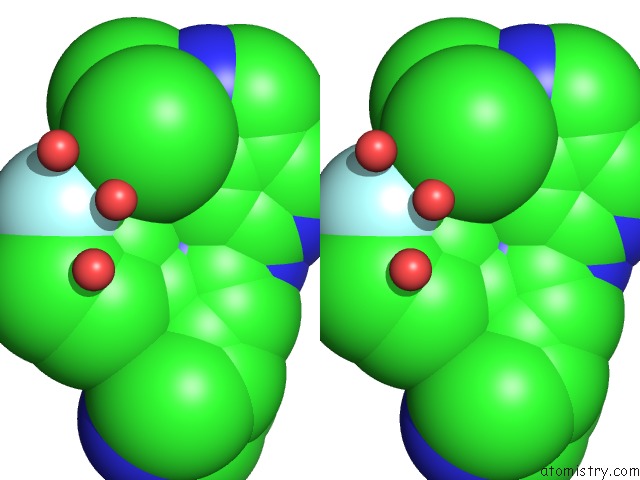
Stereo pair view
Mono view
Stereo pair view
A full contact list of Fluorine with other atoms in the F binding
site number 1 of Crystal Structure of HPK1 in Complex An Inhibitor G1858 within 5.0Å range:
|
Fluorine binding site 2 out of 2 in 6cqf
Go back to
Fluorine binding site 2 out
of 2 in the Crystal Structure of HPK1 in Complex An Inhibitor G1858
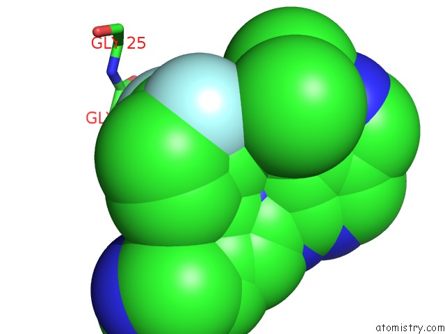
Mono view
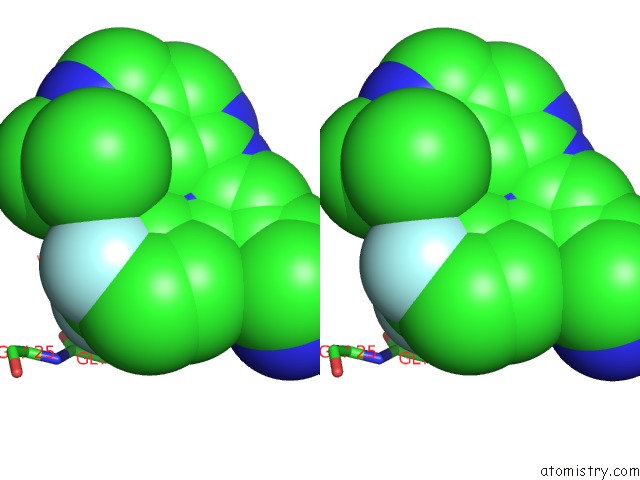
Stereo pair view
Mono view
Stereo pair view
A full contact list of Fluorine with other atoms in the F binding
site number 2 of Crystal Structure of HPK1 in Complex An Inhibitor G1858 within 5.0Å range:
|
Reference:
P.Wu,
C.J.Sneeringer,
K.E.Pitts,
E.S.Day,
B.K.Chan,
B.Wei,
I.Lehoux,
K.Mortara,
H.Li,
J.Wu,
Y.Franke,
J.G.Moffat,
J.L.Grogan,
T.P.Heffron,
W.Wang.
Hematopoietic Progenitor Kinase-1 Structure in A Domain-Swapped Dimer. Structure V. 27 125 2019.
ISSN: ISSN 1878-4186
PubMed: 30503777
DOI: 10.1016/J.STR.2018.10.025
Page generated: Tue Jul 15 10:27:41 2025
ISSN: ISSN 1878-4186
PubMed: 30503777
DOI: 10.1016/J.STR.2018.10.025
Last articles
F in 6QEEF in 6QEI
F in 6QEJ
F in 6QEF
F in 6QBS
F in 6QED
F in 6QE1
F in 6QAS
F in 6QDZ
F in 6QDY