Fluorine »
PDB 6dmg-6e69 »
6e40 »
Fluorine in PDB 6e40: Crystal Structure of the Indoleamine 2,3-Dioxygenase 1 (IDO1) in Complexed with Ferric Heme and Epacadostat
Enzymatic activity of Crystal Structure of the Indoleamine 2,3-Dioxygenase 1 (IDO1) in Complexed with Ferric Heme and Epacadostat
All present enzymatic activity of Crystal Structure of the Indoleamine 2,3-Dioxygenase 1 (IDO1) in Complexed with Ferric Heme and Epacadostat:
1.13.11.52;
1.13.11.52;
Protein crystallography data
The structure of Crystal Structure of the Indoleamine 2,3-Dioxygenase 1 (IDO1) in Complexed with Ferric Heme and Epacadostat, PDB code: 6e40
was solved by
S.Luo,
L.Tong,
with X-Ray Crystallography technique. A brief refinement statistics is given in the table below:
| Resolution Low / High (Å) | 46.86 / 2.31 |
| Space group | P 21 21 2 |
| Cell size a, b, c (Å), α, β, γ (°) | 80.367, 200.144, 114.741, 90.00, 90.00, 90.00 |
| R / Rfree (%) | 20.8 / 25.4 |
Other elements in 6e40:
The structure of Crystal Structure of the Indoleamine 2,3-Dioxygenase 1 (IDO1) in Complexed with Ferric Heme and Epacadostat also contains other interesting chemical elements:
| Bromine | (Br) | 3 atoms |
| Iron | (Fe) | 4 atoms |
Fluorine Binding Sites:
The binding sites of Fluorine atom in the Crystal Structure of the Indoleamine 2,3-Dioxygenase 1 (IDO1) in Complexed with Ferric Heme and Epacadostat
(pdb code 6e40). This binding sites where shown within
5.0 Angstroms radius around Fluorine atom.
In total 3 binding sites of Fluorine where determined in the Crystal Structure of the Indoleamine 2,3-Dioxygenase 1 (IDO1) in Complexed with Ferric Heme and Epacadostat, PDB code: 6e40:
Jump to Fluorine binding site number: 1; 2; 3;
In total 3 binding sites of Fluorine where determined in the Crystal Structure of the Indoleamine 2,3-Dioxygenase 1 (IDO1) in Complexed with Ferric Heme and Epacadostat, PDB code: 6e40:
Jump to Fluorine binding site number: 1; 2; 3;
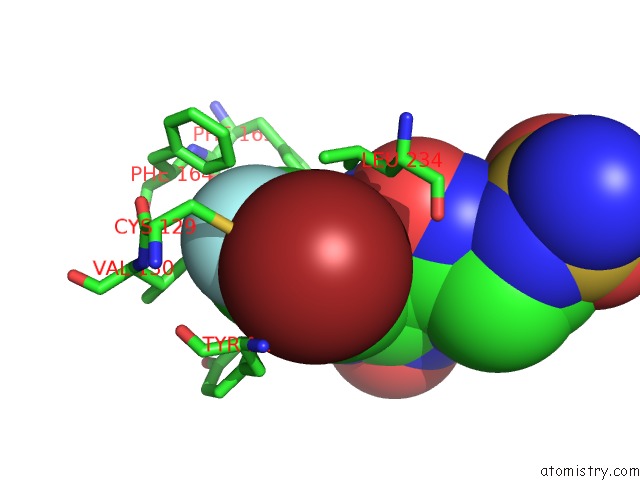
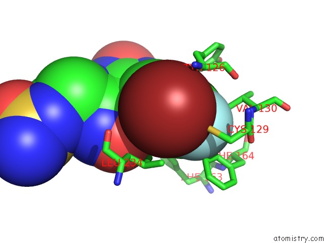
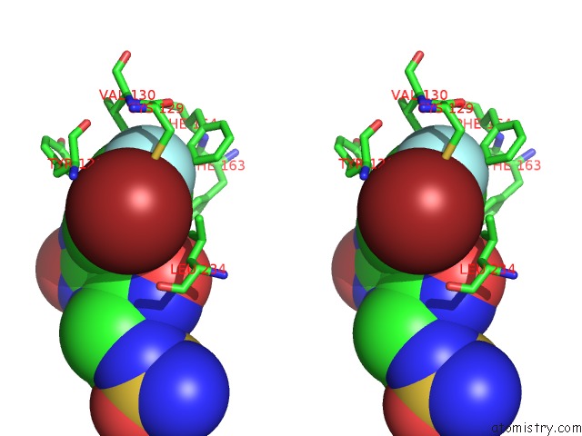
Fluorine binding site 1 out of 3 in 6e40
Go back to
Fluorine binding site 1 out
of 3 in the Crystal Structure of the Indoleamine 2,3-Dioxygenase 1 (IDO1) in Complexed with Ferric Heme and Epacadostat
Mono view
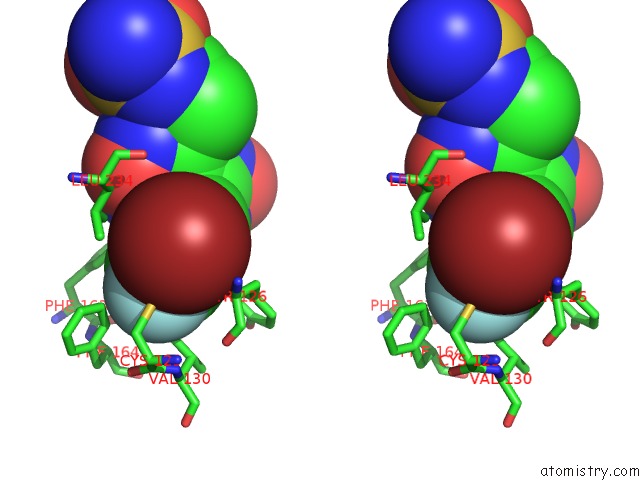
Stereo pair view
Mono view
Stereo pair view
A full contact list of Fluorine with other atoms in the F binding
site number 1 of Crystal Structure of the Indoleamine 2,3-Dioxygenase 1 (IDO1) in Complexed with Ferric Heme and Epacadostat within 5.0Å range:
|
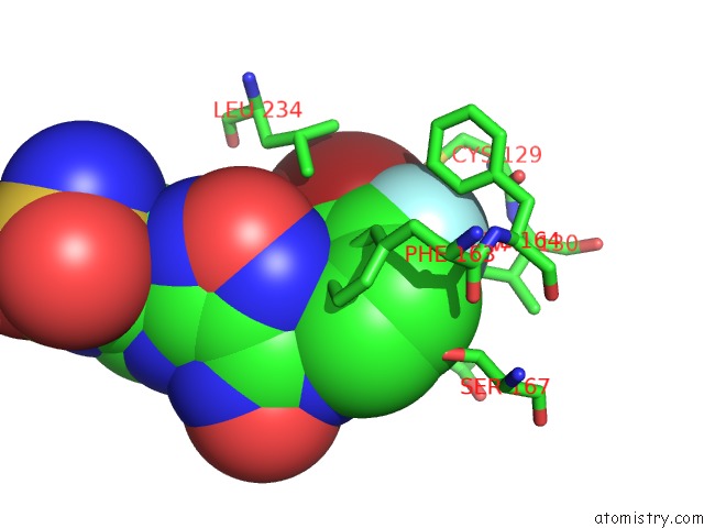
Fluorine binding site 2 out of 3 in 6e40
Go back to
Fluorine binding site 2 out
of 3 in the Crystal Structure of the Indoleamine 2,3-Dioxygenase 1 (IDO1) in Complexed with Ferric Heme and Epacadostat
Mono view
Stereo pair view
Mono view
Stereo pair view
A full contact list of Fluorine with other atoms in the F binding
site number 2 of Crystal Structure of the Indoleamine 2,3-Dioxygenase 1 (IDO1) in Complexed with Ferric Heme and Epacadostat within 5.0Å range:
|
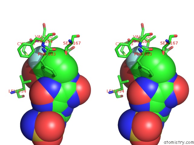
Fluorine binding site 3 out of 3 in 6e40
Go back to
Fluorine binding site 3 out
of 3 in the Crystal Structure of the Indoleamine 2,3-Dioxygenase 1 (IDO1) in Complexed with Ferric Heme and Epacadostat
Mono view
Stereo pair view
Mono view
Stereo pair view
A full contact list of Fluorine with other atoms in the F binding
site number 3 of Crystal Structure of the Indoleamine 2,3-Dioxygenase 1 (IDO1) in Complexed with Ferric Heme and Epacadostat within 5.0Å range:
|
Reference:
S.Luo,
K.Xu,
S.Xiang,
J.Chen,
C.Chen,
C.Guo,
Y.Tong,
L.Tong.
High-Resolution Structures of Inhibitor Complexes of Human Indoleamine 2,3-Dioxygenase 1 in A New Crystal Form. Acta Crystallogr F Struct V. 74 717 2018BIOL Commun.
ISSN: ESSN 2053-230X
PubMed: 30387777
DOI: 10.1107/S2053230X18012955
Page generated: Tue Jul 15 10:58:22 2025
ISSN: ESSN 2053-230X
PubMed: 30387777
DOI: 10.1107/S2053230X18012955
Last articles
F in 6ZGMF in 6ZBT
F in 6ZG9
F in 6ZFU
F in 6ZFT
F in 6ZDE
F in 6ZFS
F in 6ZDI
F in 6ZCU
F in 6ZCR