Fluorine »
PDB 6ony-6p3u »
6p3u »
Fluorine in PDB 6p3u: Crystal Structure of Eis From Mycobacterium Tuberculosis in Complex with Inhibitor SGT335
Protein crystallography data
The structure of Crystal Structure of Eis From Mycobacterium Tuberculosis in Complex with Inhibitor SGT335, PDB code: 6p3u
was solved by
A.Punetha,
S.Garneau-Tsodikova,
O.V.Tsodikov,
with X-Ray Crystallography technique. A brief refinement statistics is given in the table below:
| Resolution Low / High (Å) | 35.00 / 2.55 |
| Space group | H 3 2 |
| Cell size a, b, c (Å), α, β, γ (°) | 175.441, 175.441, 122.946, 90.00, 90.00, 120.00 |
| R / Rfree (%) | 17.6 / 20.6 |
Fluorine Binding Sites:
The binding sites of Fluorine atom in the Crystal Structure of Eis From Mycobacterium Tuberculosis in Complex with Inhibitor SGT335
(pdb code 6p3u). This binding sites where shown within
5.0 Angstroms radius around Fluorine atom.
In total 2 binding sites of Fluorine where determined in the Crystal Structure of Eis From Mycobacterium Tuberculosis in Complex with Inhibitor SGT335, PDB code: 6p3u:
Jump to Fluorine binding site number: 1; 2;
In total 2 binding sites of Fluorine where determined in the Crystal Structure of Eis From Mycobacterium Tuberculosis in Complex with Inhibitor SGT335, PDB code: 6p3u:
Jump to Fluorine binding site number: 1; 2;
Fluorine binding site 1 out of 2 in 6p3u
Go back to
Fluorine binding site 1 out
of 2 in the Crystal Structure of Eis From Mycobacterium Tuberculosis in Complex with Inhibitor SGT335
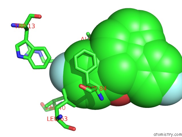
Mono view
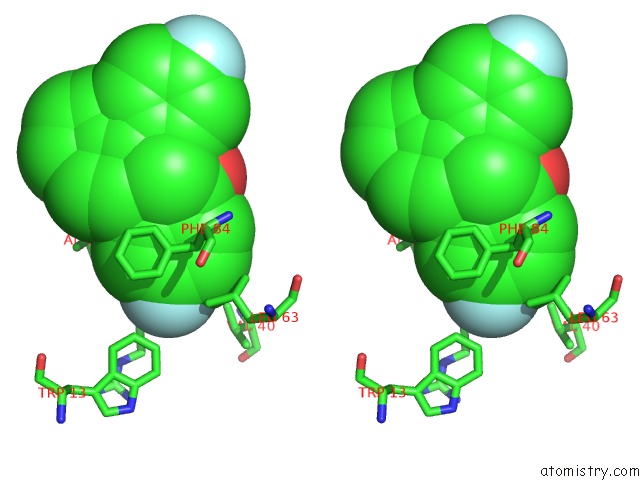
Stereo pair view
Mono view
Stereo pair view
A full contact list of Fluorine with other atoms in the F binding
site number 1 of Crystal Structure of Eis From Mycobacterium Tuberculosis in Complex with Inhibitor SGT335 within 5.0Å range:
|
Fluorine binding site 2 out of 2 in 6p3u
Go back to
Fluorine binding site 2 out
of 2 in the Crystal Structure of Eis From Mycobacterium Tuberculosis in Complex with Inhibitor SGT335
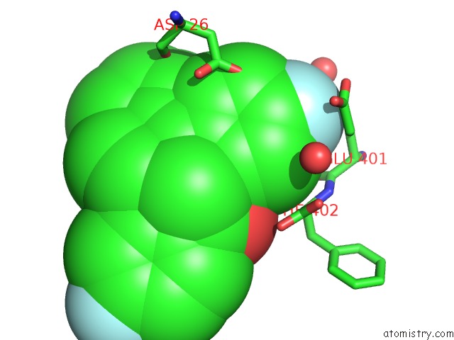
Mono view
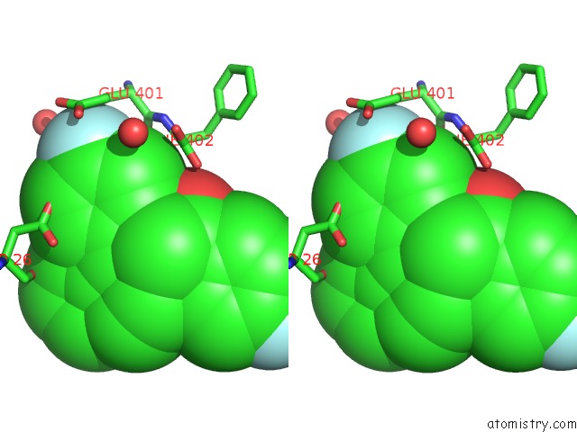
Stereo pair view
Mono view
Stereo pair view
A full contact list of Fluorine with other atoms in the F binding
site number 2 of Crystal Structure of Eis From Mycobacterium Tuberculosis in Complex with Inhibitor SGT335 within 5.0Å range:
|
Reference:
K.D.Green,
A.Punetha,
C.Hou,
S.Garneau-Tsodikova,
O.V.Tsodikov.
Probing the Robustness of Inhibitors of Tuberculosis Aminoglycoside Resistance Enzyme Eis By Mutagenesis. Acs Infect Dis. V. 5 1772 2019.
ISSN: ESSN 2373-8227
PubMed: 31433614
DOI: 10.1021/ACSINFECDIS.9B00228
Page generated: Tue Jul 15 14:28:19 2025
ISSN: ESSN 2373-8227
PubMed: 31433614
DOI: 10.1021/ACSINFECDIS.9B00228
Last articles
F in 7I2AF in 7I2D
F in 7HNS
F in 7HOG
F in 7HO4
F in 7HOL
F in 7HNQ
F in 7HMA
F in 7HNA
F in 7HMV