Fluorine »
PDB 1fko-1h1d »
1g4u »
Fluorine in PDB 1g4u: Crystal Structure of the Salmonella Tyrosine Phosphatase and Gtpase Activating Protein Sptp Bound to RAC1
Protein crystallography data
The structure of Crystal Structure of the Salmonella Tyrosine Phosphatase and Gtpase Activating Protein Sptp Bound to RAC1, PDB code: 1g4u
was solved by
C.E.Stebbins,
J.E.Galan,
with X-Ray Crystallography technique. A brief refinement statistics is given in the table below:
| Resolution Low / High (Å) | 50.00 / 2.30 |
| Space group | P 1 |
| Cell size a, b, c (Å), α, β, γ (°) | 56.722, 58.260, 60.324, 70.03, 69.51, 65.03 |
| R / Rfree (%) | 20.4 / 25.8 |
Other elements in 1g4u:
The structure of Crystal Structure of the Salmonella Tyrosine Phosphatase and Gtpase Activating Protein Sptp Bound to RAC1 also contains other interesting chemical elements:
| Magnesium | (Mg) | 1 atom |
| Aluminium | (Al) | 1 atom |
Fluorine Binding Sites:
The binding sites of Fluorine atom in the Crystal Structure of the Salmonella Tyrosine Phosphatase and Gtpase Activating Protein Sptp Bound to RAC1
(pdb code 1g4u). This binding sites where shown within
5.0 Angstroms radius around Fluorine atom.
In total 3 binding sites of Fluorine where determined in the Crystal Structure of the Salmonella Tyrosine Phosphatase and Gtpase Activating Protein Sptp Bound to RAC1, PDB code: 1g4u:
Jump to Fluorine binding site number: 1; 2; 3;
In total 3 binding sites of Fluorine where determined in the Crystal Structure of the Salmonella Tyrosine Phosphatase and Gtpase Activating Protein Sptp Bound to RAC1, PDB code: 1g4u:
Jump to Fluorine binding site number: 1; 2; 3;
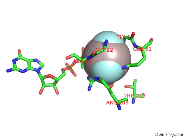
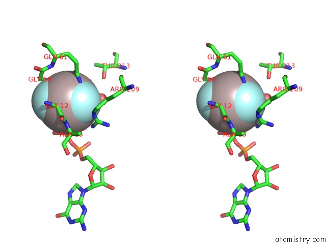
Fluorine binding site 1 out of 3 in 1g4u
Go back to
Fluorine binding site 1 out
of 3 in the Crystal Structure of the Salmonella Tyrosine Phosphatase and Gtpase Activating Protein Sptp Bound to RAC1
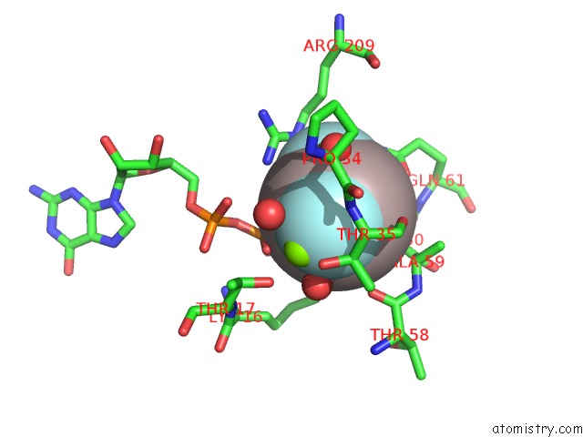
Mono view
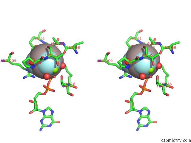
Stereo pair view
Mono view
Stereo pair view
A full contact list of Fluorine with other atoms in the F binding
site number 1 of Crystal Structure of the Salmonella Tyrosine Phosphatase and Gtpase Activating Protein Sptp Bound to RAC1 within 5.0Å range:
|
Fluorine binding site 2 out of 3 in 1g4u
Go back to
Fluorine binding site 2 out
of 3 in the Crystal Structure of the Salmonella Tyrosine Phosphatase and Gtpase Activating Protein Sptp Bound to RAC1
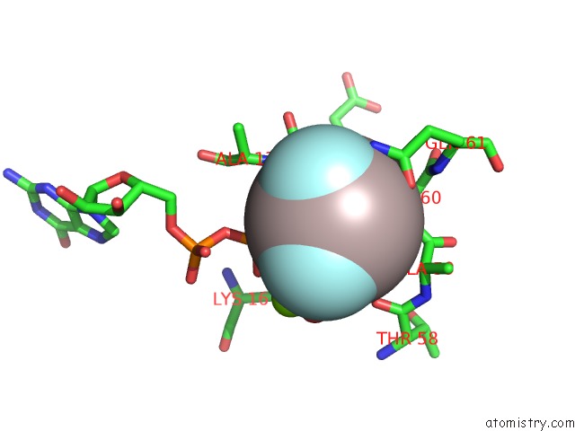
Mono view
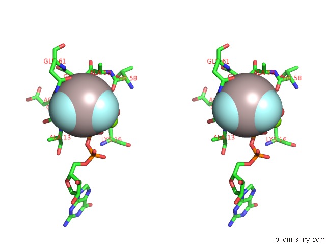
Stereo pair view
Mono view
Stereo pair view
A full contact list of Fluorine with other atoms in the F binding
site number 2 of Crystal Structure of the Salmonella Tyrosine Phosphatase and Gtpase Activating Protein Sptp Bound to RAC1 within 5.0Å range:
|
Fluorine binding site 3 out of 3 in 1g4u
Go back to
Fluorine binding site 3 out
of 3 in the Crystal Structure of the Salmonella Tyrosine Phosphatase and Gtpase Activating Protein Sptp Bound to RAC1
Mono view
Stereo pair view
Mono view
Stereo pair view
A full contact list of Fluorine with other atoms in the F binding
site number 3 of Crystal Structure of the Salmonella Tyrosine Phosphatase and Gtpase Activating Protein Sptp Bound to RAC1 within 5.0Å range:
|
Reference:
C.E.Stebbins,
J.E.Galan.
Modulation of Host Signaling By A Bacterial Mimic: Structure of the Salmonella Effector Sptp Bound to RAC1. Mol.Cell V. 6 1449 2000.
ISSN: ISSN 1097-2765
PubMed: 11163217
DOI: 10.1016/S1097-2765(00)00141-6
Page generated: Mon Jul 14 10:46:48 2025
ISSN: ISSN 1097-2765
PubMed: 11163217
DOI: 10.1016/S1097-2765(00)00141-6
Last articles
F in 4BBJF in 4BDY
F in 4BCD
F in 4BB0
F in 4BAU
F in 4B84
F in 4B6S
F in 4B9E
F in 4BA0
F in 4B76