Fluorine »
PDB 2gtn-2ihj »
2i0u »
Fluorine in PDB 2i0u: Crystal Structures of Phospholipases A2 From Vipera Nikolskii Venom Revealing Triton X-100 Bound in Hydrophobic Channel
Enzymatic activity of Crystal Structures of Phospholipases A2 From Vipera Nikolskii Venom Revealing Triton X-100 Bound in Hydrophobic Channel
All present enzymatic activity of Crystal Structures of Phospholipases A2 From Vipera Nikolskii Venom Revealing Triton X-100 Bound in Hydrophobic Channel:
3.1.1.4;
3.1.1.4;
Protein crystallography data
The structure of Crystal Structures of Phospholipases A2 From Vipera Nikolskii Venom Revealing Triton X-100 Bound in Hydrophobic Channel, PDB code: 2i0u
was solved by
W.Gao,
R.C.Bi,
with X-Ray Crystallography technique. A brief refinement statistics is given in the table below:
| Resolution Low / High (Å) | 29.00 / 2.20 |
| Space group | H 3 2 |
| Cell size a, b, c (Å), α, β, γ (°) | 76.284, 76.284, 304.395, 90.00, 90.00, 120.00 |
| R / Rfree (%) | 20.9 / 23.6 |
Other elements in 2i0u:
The structure of Crystal Structures of Phospholipases A2 From Vipera Nikolskii Venom Revealing Triton X-100 Bound in Hydrophobic Channel also contains other interesting chemical elements:
| Calcium | (Ca) | 2 atoms |
Fluorine Binding Sites:
The binding sites of Fluorine atom in the Crystal Structures of Phospholipases A2 From Vipera Nikolskii Venom Revealing Triton X-100 Bound in Hydrophobic Channel
(pdb code 2i0u). This binding sites where shown within
5.0 Angstroms radius around Fluorine atom.
In total 3 binding sites of Fluorine where determined in the Crystal Structures of Phospholipases A2 From Vipera Nikolskii Venom Revealing Triton X-100 Bound in Hydrophobic Channel, PDB code: 2i0u:
Jump to Fluorine binding site number: 1; 2; 3;
In total 3 binding sites of Fluorine where determined in the Crystal Structures of Phospholipases A2 From Vipera Nikolskii Venom Revealing Triton X-100 Bound in Hydrophobic Channel, PDB code: 2i0u:
Jump to Fluorine binding site number: 1; 2; 3;
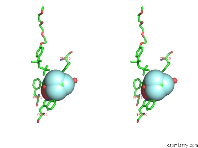
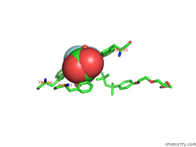
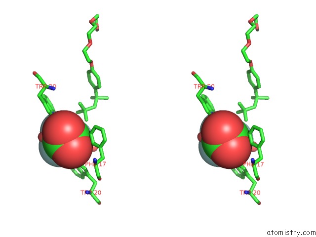
Fluorine binding site 1 out of 3 in 2i0u
Go back to
Fluorine binding site 1 out
of 3 in the Crystal Structures of Phospholipases A2 From Vipera Nikolskii Venom Revealing Triton X-100 Bound in Hydrophobic Channel
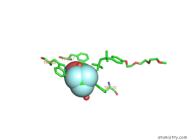
Mono view
Stereo pair view
Mono view
Stereo pair view
A full contact list of Fluorine with other atoms in the F binding
site number 1 of Crystal Structures of Phospholipases A2 From Vipera Nikolskii Venom Revealing Triton X-100 Bound in Hydrophobic Channel within 5.0Å range:
|
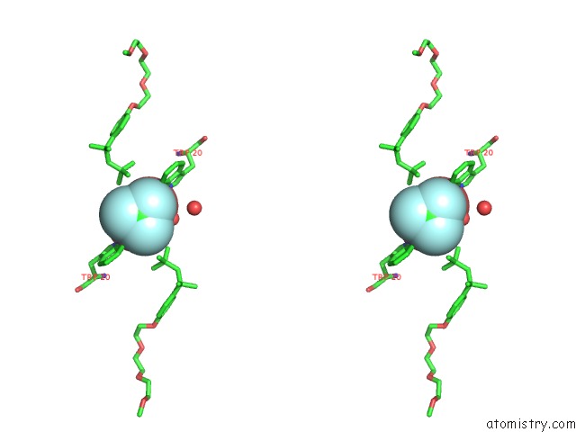
Fluorine binding site 2 out of 3 in 2i0u
Go back to
Fluorine binding site 2 out
of 3 in the Crystal Structures of Phospholipases A2 From Vipera Nikolskii Venom Revealing Triton X-100 Bound in Hydrophobic Channel
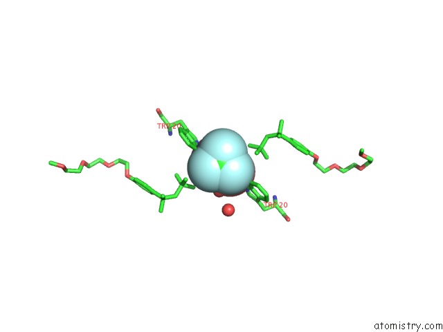
Mono view
Stereo pair view
Mono view
Stereo pair view
A full contact list of Fluorine with other atoms in the F binding
site number 2 of Crystal Structures of Phospholipases A2 From Vipera Nikolskii Venom Revealing Triton X-100 Bound in Hydrophobic Channel within 5.0Å range:
|
Fluorine binding site 3 out of 3 in 2i0u
Go back to
Fluorine binding site 3 out
of 3 in the Crystal Structures of Phospholipases A2 From Vipera Nikolskii Venom Revealing Triton X-100 Bound in Hydrophobic Channel
Mono view
Stereo pair view
Mono view
Stereo pair view
A full contact list of Fluorine with other atoms in the F binding
site number 3 of Crystal Structures of Phospholipases A2 From Vipera Nikolskii Venom Revealing Triton X-100 Bound in Hydrophobic Channel within 5.0Å range:
|
Reference:
W.Gao,
R.C.Bi.
Crystal Structures of Phospholipases A2 From Vipera Nikolskii Venom Revealing Triton X-100 Bound in Hydrophobic Channel To Be Published.
Page generated: Mon Jul 14 13:26:00 2025
Last articles
F in 7KK3F in 7KLE
F in 7KKH
F in 7KKB
F in 7KKA
F in 7KK8
F in 7KDP
F in 7KK4
F in 7KJW
F in 7KJS