Fluorine »
PDB 3rjc-3s9y »
3ruo »
Fluorine in PDB 3ruo: Complex Structure of Hevb EV93 Main Protease 3C with Rupintrivir (AG7088)
Enzymatic activity of Complex Structure of Hevb EV93 Main Protease 3C with Rupintrivir (AG7088)
All present enzymatic activity of Complex Structure of Hevb EV93 Main Protease 3C with Rupintrivir (AG7088):
3.4.22.28;
3.4.22.28;
Protein crystallography data
The structure of Complex Structure of Hevb EV93 Main Protease 3C with Rupintrivir (AG7088), PDB code: 3ruo
was solved by
Z.Kaczmarska,
R.Janowski,
L.Costenaro,
B.Coutard,
H.Norder,
B.Canard,
M.Coll,
with X-Ray Crystallography technique. A brief refinement statistics is given in the table below:
| Resolution Low / High (Å) | 30.00 / 1.50 |
| Space group | P 1 21 1 |
| Cell size a, b, c (Å), α, β, γ (°) | 38.995, 63.911, 66.357, 90.00, 90.43, 90.00 |
| R / Rfree (%) | 16.6 / 19.8 |
Other elements in 3ruo:
The structure of Complex Structure of Hevb EV93 Main Protease 3C with Rupintrivir (AG7088) also contains other interesting chemical elements:
| Magnesium | (Mg) | 2 atoms |
| Chlorine | (Cl) | 1 atom |
Fluorine Binding Sites:
The binding sites of Fluorine atom in the Complex Structure of Hevb EV93 Main Protease 3C with Rupintrivir (AG7088)
(pdb code 3ruo). This binding sites where shown within
5.0 Angstroms radius around Fluorine atom.
In total 2 binding sites of Fluorine where determined in the Complex Structure of Hevb EV93 Main Protease 3C with Rupintrivir (AG7088), PDB code: 3ruo:
Jump to Fluorine binding site number: 1; 2;
In total 2 binding sites of Fluorine where determined in the Complex Structure of Hevb EV93 Main Protease 3C with Rupintrivir (AG7088), PDB code: 3ruo:
Jump to Fluorine binding site number: 1; 2;
Fluorine binding site 1 out of 2 in 3ruo
Go back to
Fluorine binding site 1 out
of 2 in the Complex Structure of Hevb EV93 Main Protease 3C with Rupintrivir (AG7088)
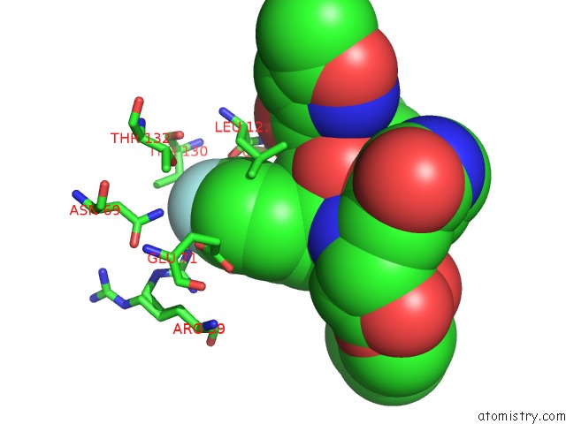
Mono view
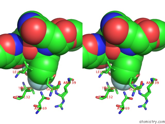
Stereo pair view
Mono view
Stereo pair view
A full contact list of Fluorine with other atoms in the F binding
site number 1 of Complex Structure of Hevb EV93 Main Protease 3C with Rupintrivir (AG7088) within 5.0Å range:
|
Fluorine binding site 2 out of 2 in 3ruo
Go back to
Fluorine binding site 2 out
of 2 in the Complex Structure of Hevb EV93 Main Protease 3C with Rupintrivir (AG7088)
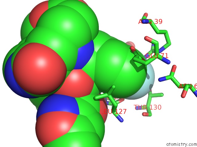
Mono view

Stereo pair view
Mono view
Stereo pair view
A full contact list of Fluorine with other atoms in the F binding
site number 2 of Complex Structure of Hevb EV93 Main Protease 3C with Rupintrivir (AG7088) within 5.0Å range:
|
Reference:
L.Costenaro,
Z.Kaczmarska,
C.Arnan,
R.Janowski,
B.Coutard,
M.Sola,
A.E.Gorbalenya,
H.Norder,
B.Canard,
M.Coll.
Structural Basis For Antiviral Inhibition of the Main Protease, 3C, From Human Enterovirus 93. J.Virol. V. 85 10764 2011.
ISSN: ISSN 0022-538X
PubMed: 21835784
DOI: 10.1128/JVI.05062-11
Page generated: Mon Jul 14 19:10:57 2025
ISSN: ISSN 0022-538X
PubMed: 21835784
DOI: 10.1128/JVI.05062-11
Last articles
F in 4J0BF in 4IYN
F in 4J0T
F in 4J03
F in 4J0P
F in 4IZW
F in 4IW8
F in 4IZT
F in 4IWF
F in 4IXE