Fluorine »
PDB 4rx0-4u7z »
4txn »
Fluorine in PDB 4txn: Crystal Structure of Uridine Phosphorylase From Schistosoma Mansoni in Complex with 5-Fluorouracil
Enzymatic activity of Crystal Structure of Uridine Phosphorylase From Schistosoma Mansoni in Complex with 5-Fluorouracil
All present enzymatic activity of Crystal Structure of Uridine Phosphorylase From Schistosoma Mansoni in Complex with 5-Fluorouracil:
2.4.2.3;
2.4.2.3;
Protein crystallography data
The structure of Crystal Structure of Uridine Phosphorylase From Schistosoma Mansoni in Complex with 5-Fluorouracil, PDB code: 4txn
was solved by
A.Marinho,
J.Torini,
L.Romanello,
A.Cassago,
R.Demarco,
J.Brandao-Neto,
H.M.Pereira,
with X-Ray Crystallography technique. A brief refinement statistics is given in the table below:
| Resolution Low / High (Å) | 29.02 / 2.00 |
| Space group | P 21 21 21 |
| Cell size a, b, c (Å), α, β, γ (°) | 95.643, 107.488, 115.576, 90.00, 90.00, 90.00 |
| R / Rfree (%) | 17.2 / 20.7 |
Fluorine Binding Sites:
The binding sites of Fluorine atom in the Crystal Structure of Uridine Phosphorylase From Schistosoma Mansoni in Complex with 5-Fluorouracil
(pdb code 4txn). This binding sites where shown within
5.0 Angstroms radius around Fluorine atom.
In total 4 binding sites of Fluorine where determined in the Crystal Structure of Uridine Phosphorylase From Schistosoma Mansoni in Complex with 5-Fluorouracil, PDB code: 4txn:
Jump to Fluorine binding site number: 1; 2; 3; 4;
In total 4 binding sites of Fluorine where determined in the Crystal Structure of Uridine Phosphorylase From Schistosoma Mansoni in Complex with 5-Fluorouracil, PDB code: 4txn:
Jump to Fluorine binding site number: 1; 2; 3; 4;
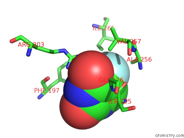
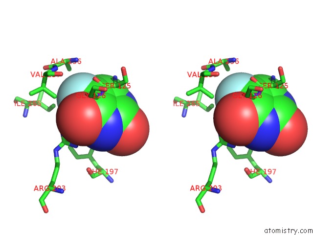
Fluorine binding site 1 out of 4 in 4txn
Go back to
Fluorine binding site 1 out
of 4 in the Crystal Structure of Uridine Phosphorylase From Schistosoma Mansoni in Complex with 5-Fluorouracil
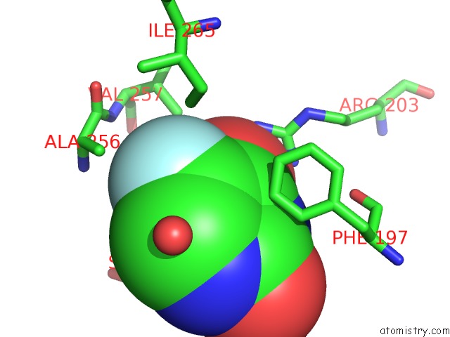
Mono view
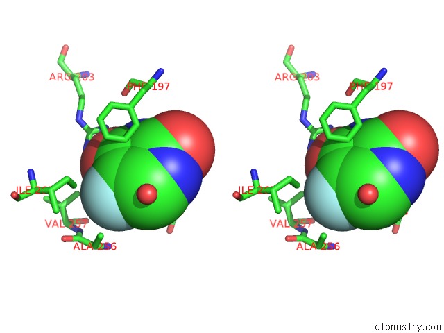
Stereo pair view
Mono view
Stereo pair view
A full contact list of Fluorine with other atoms in the F binding
site number 1 of Crystal Structure of Uridine Phosphorylase From Schistosoma Mansoni in Complex with 5-Fluorouracil within 5.0Å range:
|
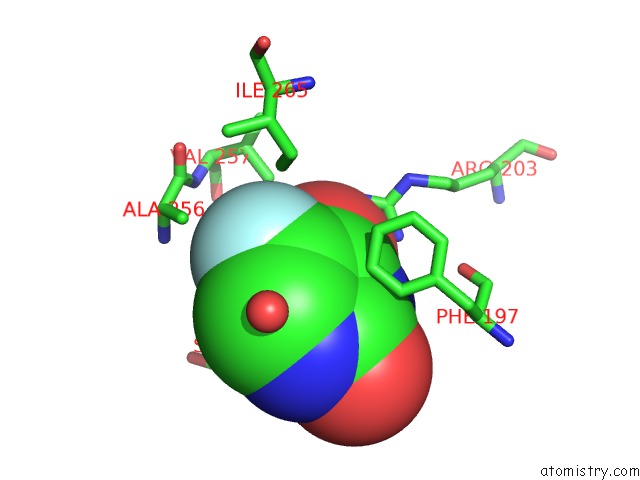
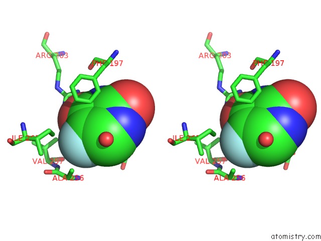
Fluorine binding site 2 out of 4 in 4txn
Go back to
Fluorine binding site 2 out
of 4 in the Crystal Structure of Uridine Phosphorylase From Schistosoma Mansoni in Complex with 5-Fluorouracil
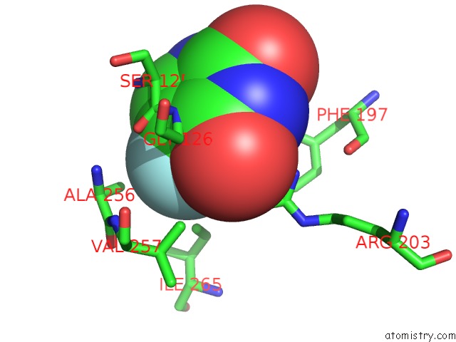
Mono view
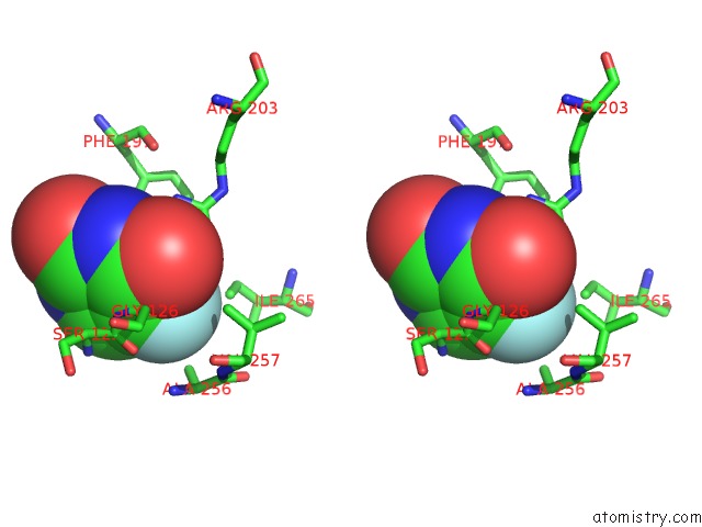
Stereo pair view
Mono view
Stereo pair view
A full contact list of Fluorine with other atoms in the F binding
site number 2 of Crystal Structure of Uridine Phosphorylase From Schistosoma Mansoni in Complex with 5-Fluorouracil within 5.0Å range:
|
Fluorine binding site 3 out of 4 in 4txn
Go back to
Fluorine binding site 3 out
of 4 in the Crystal Structure of Uridine Phosphorylase From Schistosoma Mansoni in Complex with 5-Fluorouracil
Mono view
Stereo pair view
Mono view
Stereo pair view
A full contact list of Fluorine with other atoms in the F binding
site number 3 of Crystal Structure of Uridine Phosphorylase From Schistosoma Mansoni in Complex with 5-Fluorouracil within 5.0Å range:
|
Fluorine binding site 4 out of 4 in 4txn
Go back to
Fluorine binding site 4 out
of 4 in the Crystal Structure of Uridine Phosphorylase From Schistosoma Mansoni in Complex with 5-Fluorouracil
Mono view
Stereo pair view
Mono view
Stereo pair view
A full contact list of Fluorine with other atoms in the F binding
site number 4 of Crystal Structure of Uridine Phosphorylase From Schistosoma Mansoni in Complex with 5-Fluorouracil within 5.0Å range:
|
Reference:
A.M.Da Silva Neto,
J.R.Torini De Souza,
L.Romanello,
A.Cassago,
V.H.Serrao,
R.Demarco,
J.Brandao-Neto,
R.C.Garratt,
H.D.Pereira.
Analysis of Two Schistosoma Mansoni Uridine Phosphorylases Isoforms Suggests the Emergence of A Protein with A Non-Canonical Function. Biochimie V. 125 12 2016.
ISSN: ISSN 0300-9084
PubMed: 26898674
DOI: 10.1016/J.BIOCHI.2016.02.007
Page generated: Tue Jul 15 00:46:20 2025
ISSN: ISSN 0300-9084
PubMed: 26898674
DOI: 10.1016/J.BIOCHI.2016.02.007
Last articles
F in 5JIDF in 5JN3
F in 5JMX
F in 5JMP
F in 5JLJ
F in 5JJK
F in 5JJI
F in 5JI6
F in 5JCZ
F in 5JHB