Fluorine »
PDB 5olx-5p93 »
5omg »
Fluorine in PDB 5omg: P38ALPHA in Complex with Pyrazolobenzothiazine Inhibitor COXP4M12
Enzymatic activity of P38ALPHA in Complex with Pyrazolobenzothiazine Inhibitor COXP4M12
All present enzymatic activity of P38ALPHA in Complex with Pyrazolobenzothiazine Inhibitor COXP4M12:
2.7.11.24;
2.7.11.24;
Protein crystallography data
The structure of P38ALPHA in Complex with Pyrazolobenzothiazine Inhibitor COXP4M12, PDB code: 5omg
was solved by
M.Buehrmann,
D.Rauh,
with X-Ray Crystallography technique. A brief refinement statistics is given in the table below:
| Resolution Low / High (Å) | 42.14 / 2.00 |
| Space group | P 21 21 21 |
| Cell size a, b, c (Å), α, β, γ (°) | 66.820, 74.960, 78.740, 90.00, 90.00, 90.00 |
| R / Rfree (%) | 18.2 / 24.5 |
Fluorine Binding Sites:
The binding sites of Fluorine atom in the P38ALPHA in Complex with Pyrazolobenzothiazine Inhibitor COXP4M12
(pdb code 5omg). This binding sites where shown within
5.0 Angstroms radius around Fluorine atom.
In total only one binding site of Fluorine was determined in the P38ALPHA in Complex with Pyrazolobenzothiazine Inhibitor COXP4M12, PDB code: 5omg:
In total only one binding site of Fluorine was determined in the P38ALPHA in Complex with Pyrazolobenzothiazine Inhibitor COXP4M12, PDB code: 5omg:
Fluorine binding site 1 out of 1 in 5omg
Go back to
Fluorine binding site 1 out
of 1 in the P38ALPHA in Complex with Pyrazolobenzothiazine Inhibitor COXP4M12
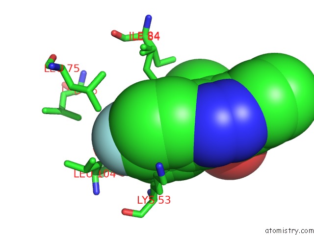
Mono view
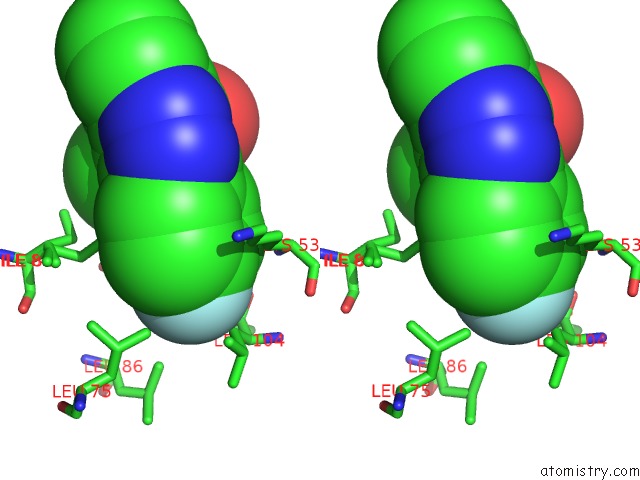
Stereo pair view
Mono view
Stereo pair view
A full contact list of Fluorine with other atoms in the F binding
site number 1 of P38ALPHA in Complex with Pyrazolobenzothiazine Inhibitor COXP4M12 within 5.0Å range:
|
Reference:
D.Bartolini,
M.Buhrmann,
M.L.Barreca,
G.Manfroni,
V.Cecchetti,
D.Rauh,
F.Galli.
Co-Crystal Structure Determination and Cellular Evaluation of 1,4-Dihydropyrazolo[4,3-C] [1,2] Benzothiazine 5,5-Dioxide P38 Alpha Mapk Inhibitors. Biochem.Biophys.Res.Commun. V. 511 579 2019.
ISSN: ESSN 1090-2104
PubMed: 30824186
DOI: 10.1016/J.BBRC.2019.02.063
Page generated: Tue Jul 15 05:51:41 2025
ISSN: ESSN 1090-2104
PubMed: 30824186
DOI: 10.1016/J.BBRC.2019.02.063
Last articles
F in 6HMRF in 6HO2
F in 6HO0
F in 6HLO
F in 6HMP
F in 6HLP
F in 6HKY
F in 6HMO
F in 6HKX
F in 6HKN