Fluorine »
PDB 6q13-6qhw »
6q6f »
Fluorine in PDB 6q6f: Crystal Structure of IDH1 R132H in Complex with HMS101
Enzymatic activity of Crystal Structure of IDH1 R132H in Complex with HMS101
All present enzymatic activity of Crystal Structure of IDH1 R132H in Complex with HMS101:
1.1.1.42;
1.1.1.42;
Protein crystallography data
The structure of Crystal Structure of IDH1 R132H in Complex with HMS101, PDB code: 6q6f
was solved by
A.Chaturvedi,
R.Goparaju,
C.Gupta,
T.Kluenemann,
M.M.Araujo Cruz,
A.Kloos,
K.Goerlich,
R.Schottmann,
E.A.Struys,
A.Ganser,
M.Preller,
M.Heuser,
with X-Ray Crystallography technique. A brief refinement statistics is given in the table below:
| Resolution Low / High (Å) | 46.09 / 3.30 |
| Space group | P 43 21 2 |
| Cell size a, b, c (Å), α, β, γ (°) | 82.396, 82.396, 301.430, 90.00, 90.00, 90.00 |
| R / Rfree (%) | 23.2 / 28.9 |
Fluorine Binding Sites:
The binding sites of Fluorine atom in the Crystal Structure of IDH1 R132H in Complex with HMS101
(pdb code 6q6f). This binding sites where shown within
5.0 Angstroms radius around Fluorine atom.
In total 2 binding sites of Fluorine where determined in the Crystal Structure of IDH1 R132H in Complex with HMS101, PDB code: 6q6f:
Jump to Fluorine binding site number: 1; 2;
In total 2 binding sites of Fluorine where determined in the Crystal Structure of IDH1 R132H in Complex with HMS101, PDB code: 6q6f:
Jump to Fluorine binding site number: 1; 2;
Fluorine binding site 1 out of 2 in 6q6f
Go back to
Fluorine binding site 1 out
of 2 in the Crystal Structure of IDH1 R132H in Complex with HMS101
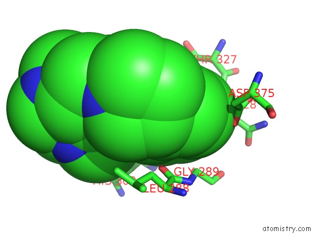
Mono view
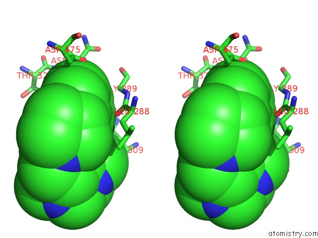
Stereo pair view
Mono view
Stereo pair view
A full contact list of Fluorine with other atoms in the F binding
site number 1 of Crystal Structure of IDH1 R132H in Complex with HMS101 within 5.0Å range:
|
Fluorine binding site 2 out of 2 in 6q6f
Go back to
Fluorine binding site 2 out
of 2 in the Crystal Structure of IDH1 R132H in Complex with HMS101
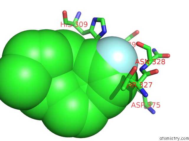
Mono view
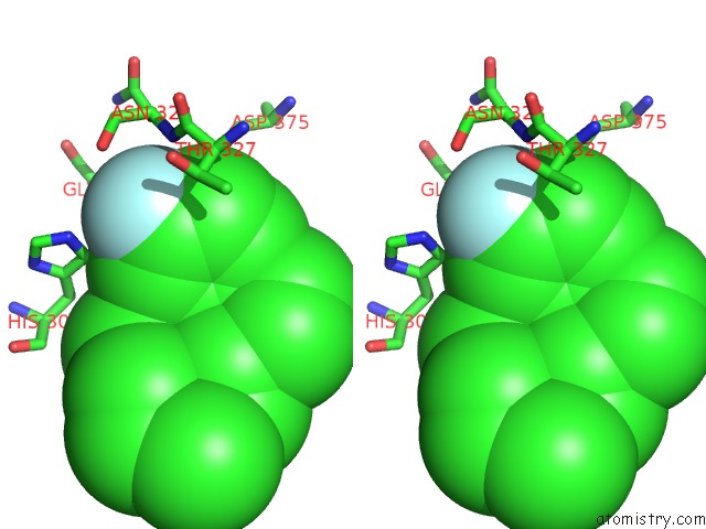
Stereo pair view
Mono view
Stereo pair view
A full contact list of Fluorine with other atoms in the F binding
site number 2 of Crystal Structure of IDH1 R132H in Complex with HMS101 within 5.0Å range:
|
Reference:
A.Chaturvedi,
R.Goparaju,
C.Gupta,
J.Weder,
T.Klunemann,
M.M.Araujo Cruz,
A.Kloos,
K.Goerlich,
R.Schottmann,
B.Othman,
E.A.Struys,
H.Bahre,
D.Grote-Koska,
K.Brand,
A.Ganser,
M.Preller,
M.Heuser.
In Vivo Efficacy of Mutant IDH1 Inhibitor Hms-101 and Structural Resolution of Distinct Binding Site. Leukemia 2019.
ISSN: ESSN 1476-5551
PubMed: 31586149
DOI: 10.1038/S41375-019-0582-X
Page generated: Tue Jul 15 14:55:16 2025
ISSN: ESSN 1476-5551
PubMed: 31586149
DOI: 10.1038/S41375-019-0582-X
Last articles
F in 7M0XF in 7M2F
F in 7M0Y
F in 7M0Z
F in 7M0M
F in 7M0V
F in 7M0W
F in 7M0U
F in 7M0N
F in 7M0T